Different intraoperative neurophysiological monitoring techniques assess the function of the brain, brainstem, spinal cord, cranial nerves, and peripheral nerves during the procedure. They are immensely valuable in the detection and prevention of neurological insult. Intraoperative monitoring is now becoming part of standard medical practices and routinely used during many surgical procedures, including the risk of neurological injury. IONM employs a wide variety of physiological principles, each with a unique application and frequently used together in the same surgery, leading to improved patient outcomes. As the benefits of monitoring become apparent, the use of different neuromonitoring techniques during the additional surgical procedure has expanded. This activity reviews the different modalities of neurophysiological monitoring, their indications, and contraindications. This activity highlights the interprofessional team's role in evaluating and improving care for patients in providing high-quality peri-operative care to detect and prevent neurologic injuries.

P/N.
Details Qty
Unit
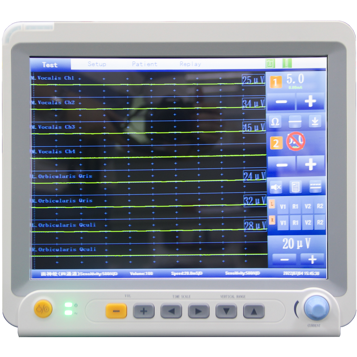
Intraoperative Neurological Monitoring Main part
Smart IOM system-main unit 1 set
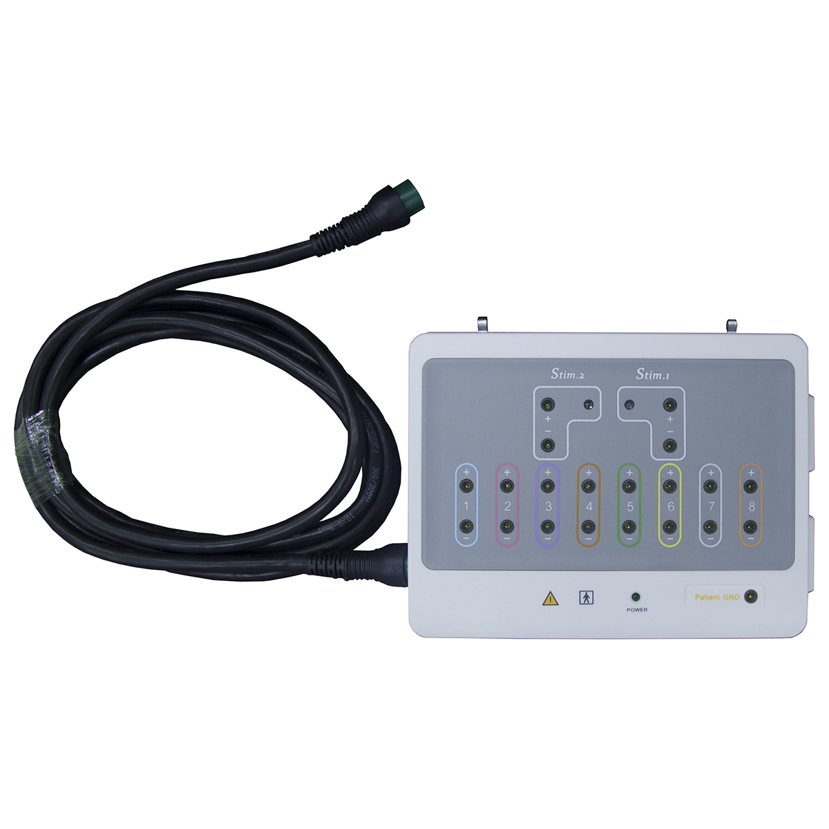
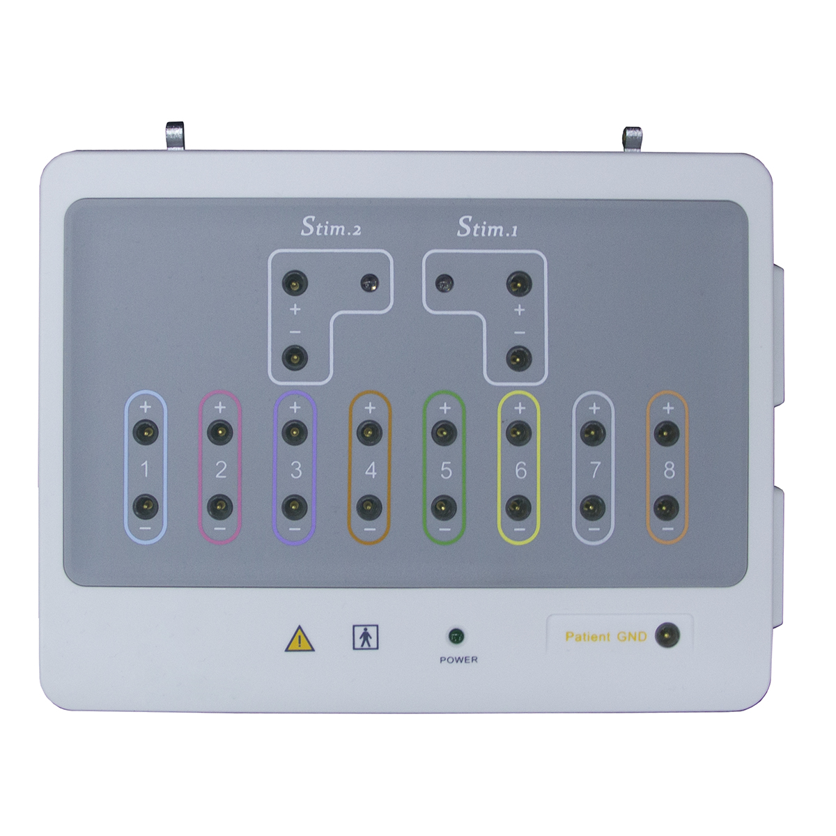
Smart IOM Communication box 1 set
Pre-amplifier 8 channels 1 set
Intraoperative Neurological Monitoring Electrode and Cable
Ground cable 1 pc
Connection cable 1 pc
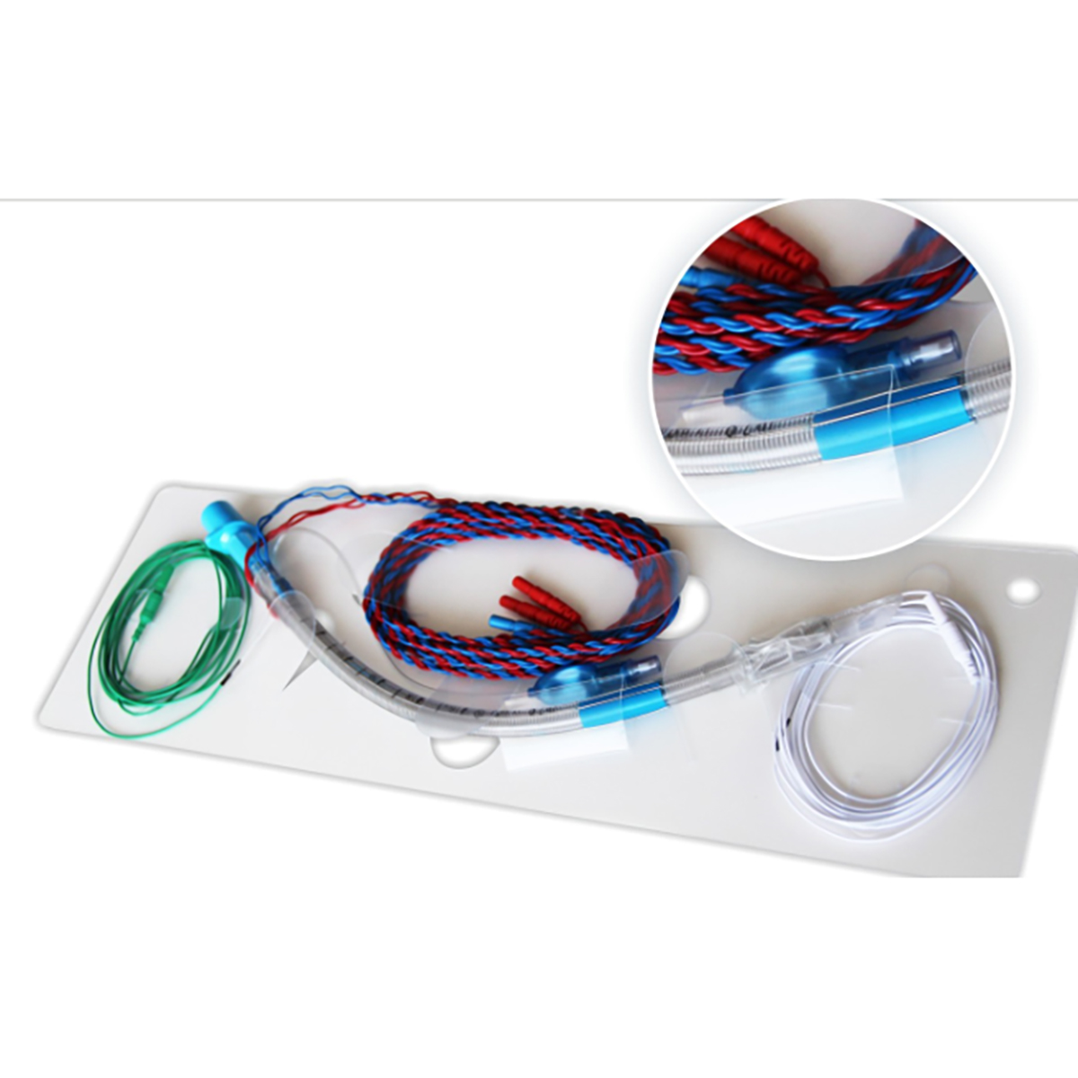
EMG endotracheal tube(Size: I.D. 7.0mm) 1 pc
Single subdermal needles 1 pc
Twisted paired subdermal needles 1 pc
Monopolar stimulation probe 1 pc
Intraoperative Neurological Monitoring Documents
Quality certificate 1 pc
Operation Manual (English version) 1 pc
EMG endotracheal tube manual 1 pc
Warranty card 1 pc
Intraoperative Neurological Monitoring Software
SmartIOM Software 1 set
Objectives:
Identify the indications for intraoperative neurophysiological monitoring.
Describe the different techniques of intraoperative neurophysiological monitoring.
Explain the importance of intraoperative neurophysiological monitoring to detect and prevent neurological injury.
Review the importance of effective communication and close cooperation between interprofessional teams in providing high-quality peri-operative care to detect and prevent neurologic injuries.
Intraoperative neurophysiological monitoring (IONM) helps assess the integrity of neural structures and consciousness during surgical procedures. It includes both continuous monitoring of neural tissue as well as the localization of vital neural structures. The goal of IONM is to identify intraoperative neural insults that allow early intervention to eliminate or to significantly minimize irreversible damage to the neurological structure and prevent a postoperative neurologic deficit. The use of neurophysiological monitoring during surgical procedures requires specific anesthesia techniques to avoid interference and signal alteration due to anesthesia.
Different modalities of intraoperative neurophysiological monitoring (IONM) are available, each monitors a specific neural pathway, and they are:
Evoked potentials including somatosensory evoked potential (SSEP), motor evoked potential (MEP), brainstem auditory evoked potential (BAEP), visual evoked potential (VEP)
Electroencephalography (EEG)
Electromyography (EMG)
Multimodal intraoperative neuromonitoring (IONM) is recommended as an effective way to avoid permanent neurologic injury during surgical procedures.[1]
Each technique of intraoperative neurophysiological monitoring monitors a specific neural pathway.
Somatosensory Evoked Potential (SSEP): SSEP monitors the dorsal column–medial lemniscus pathway, which mediates tactile discrimination, vibration, and proprioception.[2] Stimulation of sensory receptors in the skin initiates peripheral sensory nerves, which extend through the nerve root to the ipsilateral dorsal root ganglia at spinal levels. The projections from these first-order neurons form fasciculi gracilis and cuneatus, which carry impulse from the lower and upper extremities, respectively. The first synapse occurs in the lower medulla, then the impulses cross over at the level of the brainstem and form medial lemniscus. The impulse then ascends to the contralateral thalamus and relay information to the primary sensory cortex in the parietal lobe. In the upper extremities, the median and ulnar nerve are monitored, whereas, in the lower extremities, the posterior tibial and peroneal nerve are monitored.
Motor Evoked Potential (MEP): MEP monitor motor pathways, transcranial electrical stimulation elicits excitation of corticospinal projections at multiple levels. Depending on the intensity of stimulation and the placement of electrode, motor evoked potentials are generated at different levels of the brain, including superficial white mater just underneath the motor cortex, the deep white matter of the internal capsule, and pyramidal decussation. The electrical potential is recorded at the spinal cord or muscles. MEP is generated and transported via the pyramidal tract.
Visual Evoked Potential (VEP): VEP measures the functional integrity of the optic pathways from the retina to the brain's visual cortex in response to light stimulus. Visual stimulus is converted into nerve signals in the retina. These signals are transmitted via the optic pathway to the brain, from the retina to the optic nerve, optic chiasma, optic tract, lateral geniculate body, optic radiation, and visual cortex occipital lobe.
Brainstem Auditory Evoked Potential (BAEP): BAEP monitors the function of the auditory nerve and auditory pathways in the brainstem. The auditory signal travels from the cochlear hair cell to the primary auditory cortex via the vestibulocochlear nerve, superior olivary complex, lateral lemniscus, inferior colliculus, and medial geniculate body.
Electromyography (EMG): EMG monitors somatic efferent nerve activity and evaluates the functional integrity of individual nerves. EMG monitors intracranial, spinal, and peripheral nerves during surgeries. Depolarization of a motor nerve produces electrical potential within the muscles innervated by that specific nerve, and the generated electrical activity is monitored using subdermal or intramuscular electrodes.
Electroencephalography (EEG): The electrical activity measured by EEG is generated by groups of pyramidal neurons, which has cell bodies in the 3rd and 5th layer of the cerebral cortex.[6]
The intraoperative neurophysiological monitoring (IONM) systems are equipped with all the channels to monitor different modalities of IONM needed for the specific type of surgery. The monitoring system displays continuous trends and raw signals, as well as it can also trigger the stimulation of specific muscle and nerve (peripheral, cranial) and records the signal. The system also records waveforms' time/date, detail waveform data, and the technologist's notes. The system usually has a provision for rejection of artifacts related to electrocautery.


 Price is 8-20% Lower Than Other




![{pr0int $v['title']/}](https://medicalequipment-msl.com/upload/img/20141201/201412011114124238.jpg.jpg)
![{pr0int $v['title']/}](https://medicalequipment-msl.com/upload/img/20141121/201411211505023940.jpg.jpg)
![{pr0int $v['title']/}](https://medicalequipment-msl.com/upload/img/20180118/201801180944127389.jpg.jpg)
![{pr0int $v['title']/}](https://medicalequipment-msl.com/upload/img/20161011/201610111637086690.jpg.jpg)
![{pr0int $v['title']/}](https://medicalequipment-msl.com/upload/img/20180118/20180118105526596.jpg.jpg)
![{pr0int $v['title']/}](https://medicalequipment-msl.com/upload/img/20161011/201610111658196300.jpg.jpg)
![{pr0int $v['title']/}](https://medicalequipment-msl.com/upload/img/20130717/201307171206452148.jpg.jpg)
![{pr0int $v['title']/}](https://medicalequipment-msl.com/upload/img/20180118/201801181036041384.jpg.jpg)
![{pr0int $v['title']/}](https://medicalequipment-msl.com/upload/img/20170110/201701101710103968.jpg.jpg)
![{pr0int $v['title']/}](https://medicalequipment-msl.com/upload/img/20180118/201801181030223690.jpg.jpg)
![{pr0int $v['title']/}](https://medicalequipment-msl.com/upload/img/20160125/201601251803268582.jpg.jpg)
![{pr0int $v['title']/}](https://medicalequipment-msl.com/upload/img/20180118/201801181005566846.jpg.jpg)
![{pr0int $v['title']/}](https://medicalequipment-msl.com/upload/img/20180118/201801181047088940.jpg.jpg)
![{pr0int $v['title']/}](https://medicalequipment-msl.com/upload/img/20161011/201610111707201967.jpg.jpg)
![{pr0int $v['title']/}](https://medicalequipment-msl.com/upload/img/20161011/201610111614296677.jpg.jpg)
![{pr0int $v['title']/}](https://medicalequipment-msl.com/upload/img/20161011/201610111624484663.jpg.jpg)
![{pr0int $v['title']/}](https://medicalequipment-msl.com/upload/img/20130727/201307271701222050.jpg.jpg)
![{pr0int $v['title']/}](https://medicalequipment-msl.com/upload/img/20190614/201906141354244381.jpg.jpg)
![{pr0int $v['title']/}](https://medicalequipment-msl.com/upload/img/20130717/201307171144589419.jpg.jpg)
![{pr0int $v['title']/}](https://medicalequipment-msl.com/upload/img/20180118/201801180935261077.jpg.jpg)
![{pr0int $v['title']/}](https://medicalequipment-msl.com/upload/img/20130716/201307161539357763.jpg.jpg)
![{pr0int $v['title']/}](https://medicalequipment-msl.com/upload/img/20161011/201610111547084360.jpg.jpg)
![{pr0int $v['title']/}](https://medicalequipment-msl.com/upload/img/20170110/201701101734311172.jpg.jpg)
![{pr0int $v['title']/}](https://medicalequipment-msl.com/upload/img/20170110/201701101813419783.jpg.jpg)
![{pr0int $v['title']/}](https://medicalequipment-msl.com/upload/img/20170110/201701101824147215.jpg.jpg)


