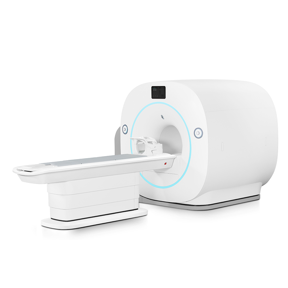
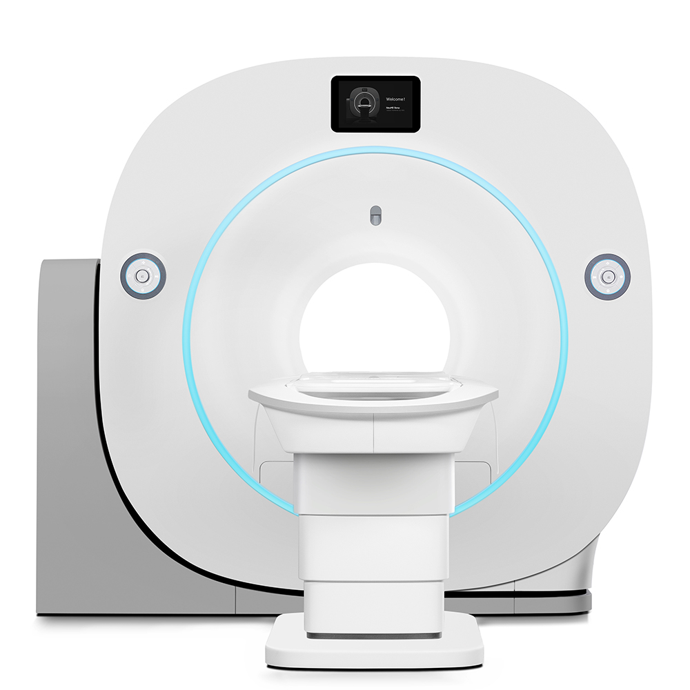
MSLMRI08 0.4T Open Permanent Magnet MRI System - Technical Specifications & Features
As a high-performance 0.4T permanent magnet MRI system, MSLMRI08 integrates advanced digital technology and clinical applicability, making it ideal for hospital-grade clinical imaging and scientific research collaboration. Below are the detailed technical parameters and core features:
1. Magnet Specifications
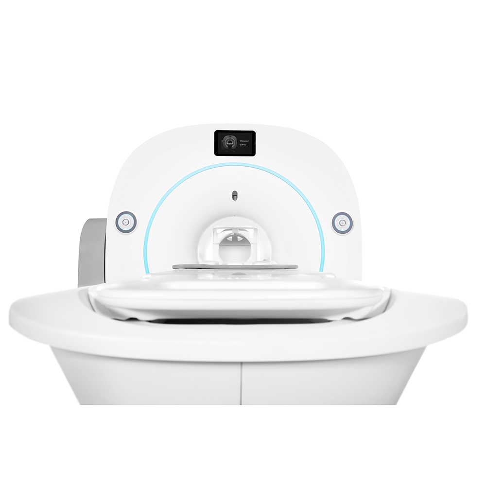
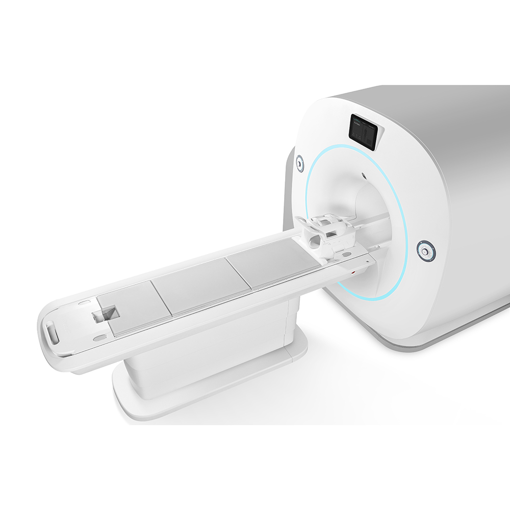
- Magnet Type: Open permanent magnet (NdFeB – Neodymium, Iron & Boron), eddy current free, open C type or 4-column type with self-temperature control
- Field Strength: 0.4T±3% (passive shimming)
- Field Homogeneity: ≤40ppm (Vpp) / ≤5ppm (Vrms) within 40×40×38cm sphere
- Vertical Gap Distance: 400mm±20mm
2. Gradient System
- Gradient Type: Full-digital gradient chain with embedded cooling design and multi-point real-time temperature control
- Gradient Strength: Super-high 46 mT/m
- Gradient Module: Digital gradient waveform generator positioned in front of gradient amplifier, realizing fiber-optic digital transmission (ensures high bandwidth and output fidelity)
- Slew Rate: Optimized for rapid imaging sequences
- Linearity: <5% within 40cm×40cm×38cm ellipsoid
- Cooling System: Wind cooling for gradient power; no additional cooling required for gradient coil
3. Spectrometer
- Type: Boundless Platform Fiber Distributed Spectrometer
- Core Advantages: Stronger real-time performance, higher SNR (Signal-to-Noise Ratio), wider scalability, broader openness
- Function: Supports fully open scientific research platform and provides scientific research collaboration solutions
4. Imaging Technology & Performance
- Acceleration Technology: DeepR-AI acceleration technology (ensures ultra-fast scanning speed and fidelity)
- High-Resolution Imaging: VAST technology enables 3D isotropic high-resolution imaging for T1, T2, FLAIR and PD sequences, supporting distortion-free MPR reconstruction (applicable to various body parts)
- DWI Performance: MUSIC-HDDWI technology with high spatial resolution and clear anatomical details; supports b values up to 10,000 (adapts to high-performance imaging requirements)
- Temporal Resolution: 4 sec/phase (achieved via DANCE K-space dynamic random sparse sampling)
- Spatial Resolution: 0.7mm×0.7mm×2.5mm (for multi-phase dynamic enhancement scanning)
- Scan Efficiency: 45% reduction in scanning time for brain imaging (compared to conventional techniques) with consistent image quality
- Quantitative Imaging: Supports precise quantification via "Six Echoes, Six Contrasts, Precise Quant, One Acquisition" mode
5. RF System
- Transmitter & Receiver: Digital type (imported components), multi-channel configuration
- RF Coils: Phase Array (PA) RF receiving coils with intelligent identification (standard configurations include Head, Body (large), Body (small), Neck, Knee, Breast, Wrist coils)
- Frequency Range: DC-22MHz
- Maximum Receiver Bandwidth: 1.25MHz
- Pre-amplifier Gain: Up to 30dB
- Noise Figure: 0.3dB
- RF Amplifier: ≥6kw (17.00MHz, imported components)
- Auto Functions: Auto RF correction, auto frequency lock, auto gain adjusting, auto coil identification, auto coil tuning, automatic shimming linear compensation
6. Software & Workstation
- Operating System: English interface, NeuUI 2.0 Dual Screen Design (more convenient and professional)
- Post-Processing Software: Supports advanced functions including SWI, tSWI, PDMAP, T1MAP, R2*MAP, QSM, ADC, DTI, ASL, MRS, MRCP
- AI Technology: NeuAI-Denoise Dual-domain fidelity (ensures data authenticity and image stability during reconstruction)
- Quantitative Analysis Tools: LiverQuant (for hepatic fat quantification and iron deposition analysis), KneeQuant (non-invasive evaluation of cartilage/meniscus biochemical changes in early osteoarthritis)
7. Hardware Configuration
- Computer System: 2nd generation Intel Core™ (≥3.3GHz), 4GB memory, 500GB hard drive, 1GB graphics memory
- Monitor: 21” TFT monitor with ≥1280×1024 resolution
- Removable Storage: DVD+RW double burn
- Patient System: Examination table, intercom system, background music system
8. Application Scope
Comprehensive clinical applications covering:
- Nerves and Blood Vessels (e.g., brachial plexus imaging)
- Joints (knee, wrist, etc.)
- Abdomen
- Total Spine & PET-LIKE imaging
- Other body parts (head, neck, breast, etc.)
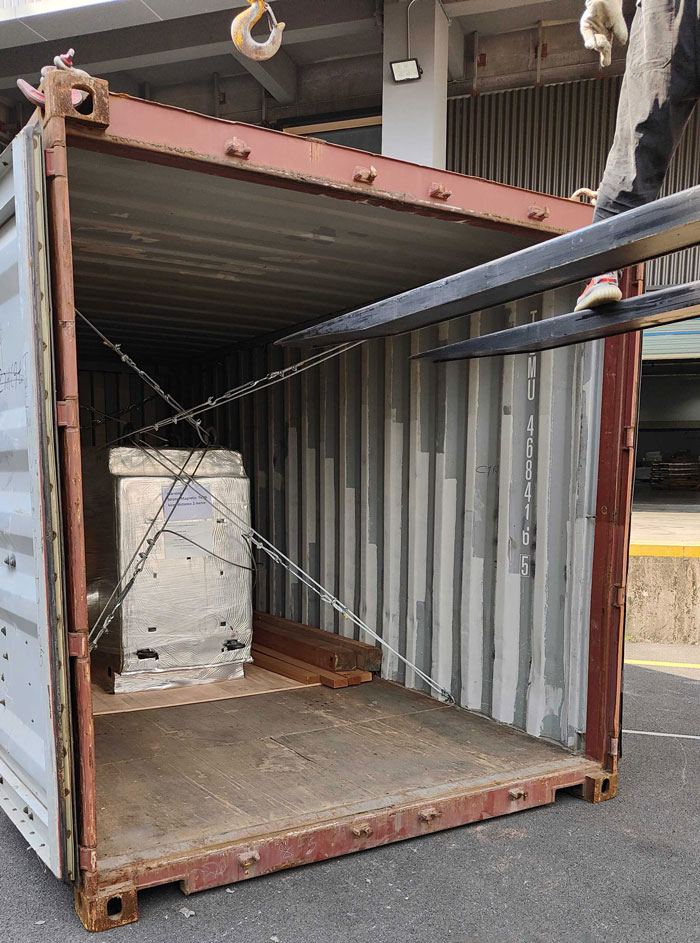
9. After-sales & Logistics
- Global Service Network: HQ service center, training centers, and spare parts centers worldwide
- Remote Service: Real-time remote technical support for immediate problem-solving
- Logistics Support: Fast response for spare parts and supplies via global logistics network
- Downtime Minimization: Proactive and prompt fault identification and correction


 Price is 8-20% Lower Than Other