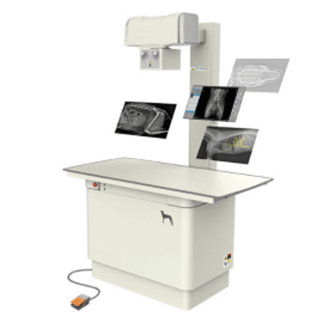
Simple And Convenient Digital Veterinary X-Ray Machine MSLVX28
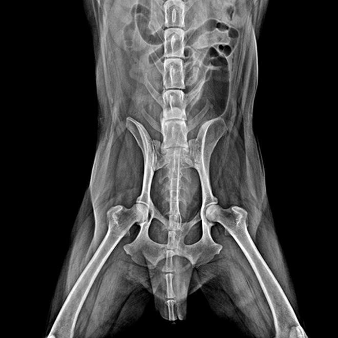
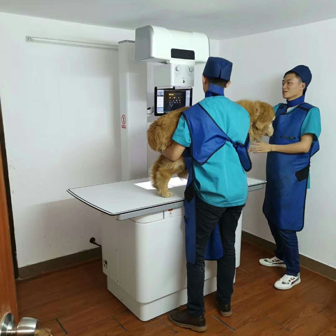
- DR for pets
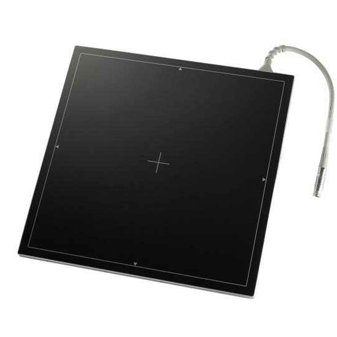
- Plat panel: Rayence1717SCCMaging size: 43 cm × 43 cm
- Select mirror and rotate at any angle according to different positions
- Display patient information / examination information / hospital information / image information
- Patient examination report form edit, save, printout function
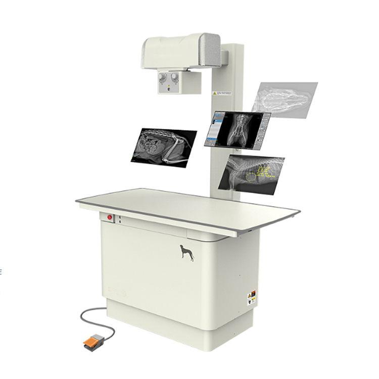
Digital Veterinary X-Ray Machine MSLVX28 Technical parameters:
·Plat panel(Rayence1717SCC)
·maging size: 43 cm × 43 cm
·Detector type: CSI
·Pixel pitch: 127um
·High frequency high voltage generator
·Input power: 200-240 V
·Output power: 32 kW
·MA: 320mA
·kV adjustment range: 40~150kV
·X-ray tube(Toshiba E7240)
·Tube focus size: Small focus 0.6 mm, large focus 1.2 mm
·Tube target angle: 12°
·Rotating anode speed: 3200r/min
·Pet DR dedicated photography bed (high-end four-way floating photography bed)
·Bed surface four-way motion stroke: Lateral stroke: 25cm ·Longitudinal stroke: 54cm
·Bed size: 1400mmX700mm
·The bed is equipped with a urinary catheter groove design to prevent pets from overflowing the urine and damaging the device
·Bed surface with low-ray attenuation and scratch-resistant material
·Fixed straps at both ends of the bed
·Bed height: 80cm
·Beam (Long Focal Length, Low Leakage) Bed Height
·Illumination: Illumination ≥200LUX, high illumination, easy to confirm shooting field of view
·Beamer positioning lamp delay time: 30s
·Positioning light type: LED (long life, low power consumption)
·With the function of automatic lighting when the bed surface moves, reducing labor intensity
·touch screen
·Size: 15.6 inches
·Touch method: Capacitive touch
·Exposure tips: With exposure sound prompt function
·Features: Display on the same screen, preview images near the station, switch parts, adjust parameters, etc.
·Remote exposure
·Exposure method: With compartment remote exposure remote control, support 15 meters remote exposure operation
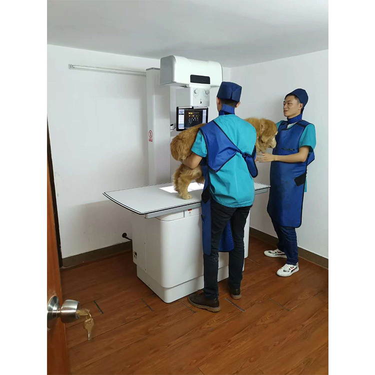
Digital Veterinary X-Ray Machine MSLVX28 Digital image acquisition and processing workstation:
·Host: DELL
·operating system: Windows 10 64bit
·CPU: intel i3 4 GHz
·RAM: 4 G
·hard disk: 1TB
·Display
·Size: 24inch
·Screen: LED
·Processing system
·Software functions
·Patient management: Includes interface to be checked, rapid emergency registration, checked patient interface
·an examination: 3D pet simulation diagram, inspection site selection, ·automatic selection of photographic parameters,
·Image review: Image thumbnail preview and other functions
·Configuration: Processing, display, layout, tools, mainly for image ·viewing and processing
·Advanced clinical functions
·Multi-band processing function: Intelligent layer processing of images, and perform optimal algorithm processing on the layered images respectively
·Various pet tissue density processing functions: Management to ensure the best image display
·High brightness denoising and enhancement: For different pet tissue density differences, targeted image algorithm parameters
·Detail enhancements: Number can get good image quality
·Extensive measurement tools for pets: Minimize image noise and effectively enhance the image to ensure the image
·Case management
·Case management functions: Management of patient information, examination information and images
·Inquiry service: DICOM3.0 standard Worklist query service interface.
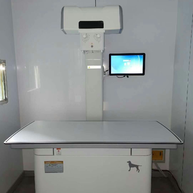
Digital Veterinary X-Ray Machine MSLVX28 Image Acquisition:
·Communication function with high voltage generator, the reference exposure parameters of each position can be directly called from the software, and the high voltage exposure can be set automatically
·Parameters, simple and convenient operation
·Real-time automatic window width and window adjustment
·Real-time edge enhancement
·Select mirror and rotate at any angle according to different positions
·Display patient information / examination information / hospital information / image information
·Real-time reminder system storage space
·Image Processing
·Window width and window adjustment
·Positive and negative film conversion, image zoom, pan, mirror, rotate, walk
·Can choose to zoom in on the area of interest, full screen display
·Image annotation capabilities including orientation and text
·Real-time automatic ROI cropping
·With advanced image processing mode: it can achieve the ideal after processing the image that is not ideal with the advanced image processing mode
·Image output interface
·DICOM3.0 standard laser camera output, you can easily select the configured solution (film size, typesetting) and print
·DICOM3.0 standard archiving service, which can archive images to the server and support automatic sending in the background
·Image backup function, backup images to CD / DVD, backup disc comes with browser to automatically play images
·Image export, patient examination can be exported to the specified location, support for JPG, PNG, DICOM and other formats of image export.
·Report output
·Patient examination report form edit, save, printout function
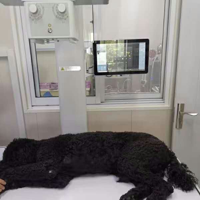


 Price is 8-20% Lower Than Other







![{pr0int $v['title']/}](https://medicalequipment-msl.com/upload/img/20150722/201507221853137106.jpg.jpg)
![{pr0int $v['title']/}](https://medicalequipment-msl.com/upload/img/20150722/201507221843206734.jpg.jpg)
![{pr0int $v['title']/}](https://medicalequipment-msl.com/upload/img/20130717/201307171850418734.jpg.jpg)
![{pr0int $v['title']/}](https://medicalequipment-msl.com/upload/img/20150722/201507221604564212.jpg.jpg)
![{pr0int $v['title']/}](https://medicalequipment-msl.com/upload/img/20130717/201307171824028797.jpg.jpg)
![{pr0int $v['title']/}](https://medicalequipment-msl.com/upload/img/20150722/201507221712547748.jpg.jpg)
![{pr0int $v['title']/}](https://medicalequipment-msl.com/upload/img/20130718/201307180947028636.jpg.jpg)
![{pr0int $v['title']/}](https://medicalequipment-msl.com/upload/img/20130718/201307181124299582.jpg.jpg)
![{pr0int $v['title']/}](https://medicalequipment-msl.com/upload/img/20150722/201507221801469794.jpg.jpg)
![{pr0int $v['title']/}](https://medicalequipment-msl.com/upload/img/20130718/201307180940488024.jpg.jpg)
![{pr0int $v['title']/}](https://medicalequipment-msl.com/upload/img/20130718/201307180928109021.jpg.jpg)
![{pr0int $v['title']/}](https://medicalequipment-msl.com/upload/img/20130717/201307171829232589.jpg.jpg)
![{pr0int $v['title']/}](https://medicalequipment-msl.com/upload/img/20150722/201507221824176080.jpg.jpg)
![{pr0int $v['title']/}](https://medicalequipment-msl.com/upload/img/20130717/201307171841265112.jpg.jpg)
![{pr0int $v['title']/}](https://medicalequipment-msl.com/upload/img/20130718/201307180954143510.jpg.jpg)
![{pr0int $v['title']/}](https://medicalequipment-msl.com/upload/img/20200330/202003301253226992.jpg.jpg)
![{pr0int $v['title']/}](https://medicalequipment-msl.com/upload/img/20200402/202004021936462479.jpg.jpg)
![{pr0int $v['title']/}](https://medicalequipment-msl.com/upload/img/20200420/202004202105173257.jpg.jpg)
![{pr0int $v['title']/}](https://medicalequipment-msl.com/upload/img/20200804/202008041015593525.jpg.jpg)


