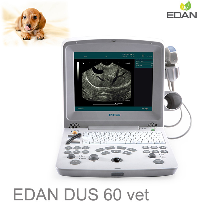
Value compact black and white portable ultrasound for OB, Gynecology, Podiatry, Orthopedic, Veterinary and applications needing PW but no Color Doppler.
Harmonic signals are produced as ultrasound waves propagate through the body. Because these signals are produced in the body, they are not influenced by artifact inducing fat near the skin surface. Consequently, an image formed using only the harmonic signal will have less clutter and can be more diagnostic.
Better than Sonoscape A6 – more features and benefits.
Compact, lightweight, easy to use, intuitive design portable ultrasound. PW for physiologic diagnostic value. Harmonics to increase image quality. A very good value portable ultrasound.
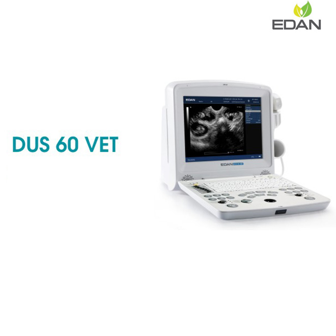
DUS60Vet Applications: DUS 60 (2013) VET is an impressive new compact ultrasound system providing superb value and best-in-class quality across a wide range of veterinary applications. With the added support of pulsed wave Doppler imaging, the DUS 60 VET meets higher diagnostic requirements.
Imageing mode: B, 2B, 4B, B+M, M, and PW
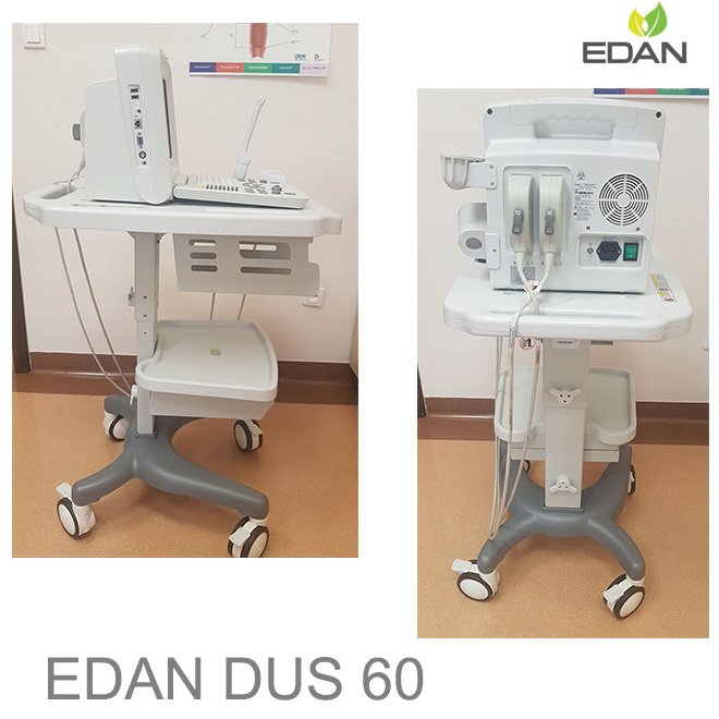
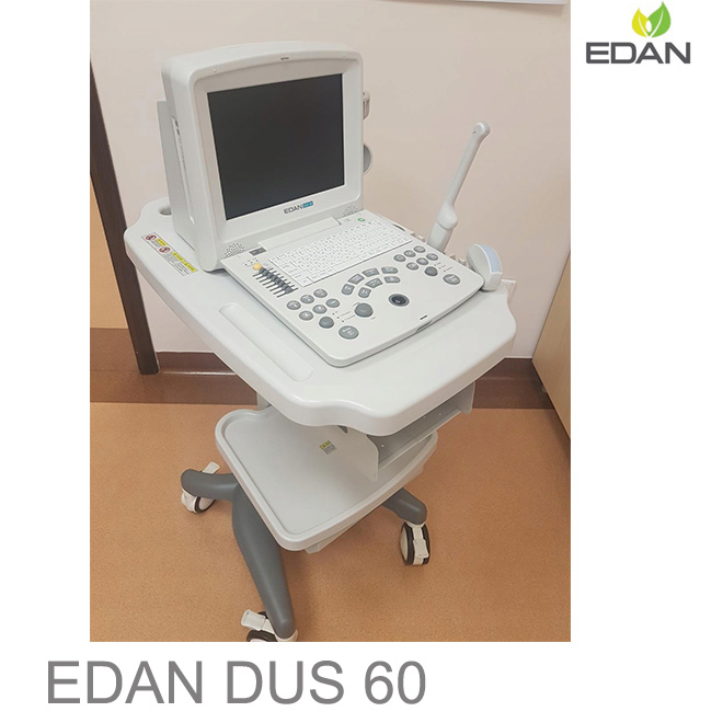
DUS 60 main unit
Two transducer connector
256-frame cine loop memory
504MB built-in image storag
Two USB ports
Pseudo color: 7 types
Measurement & Calculation software packages
Multi-purpose for abdomen, OB, GYN, Urology, Small parts and biopsy
Digital Beam former
Two transducer connectors
Multi-frequency transducer series, fulfill different applications
Maximum frequency up to 10MHz
12.1” LCD monitor
USB ports support storage, transferring and printing
THI (Tissue Harmonic Imaging)
TSI (Tissue Specific Imaging)
PW imaging
Lithium rechargeable battery
IP (Image Processing) function
Backlit keyboard
DICOM 3.0 (optional)
C343UA Convex
C362UA Convex
C321UA Micro-convex
C613UA Micro-convex
L763UA Linear
L743UA Linear
L742UA Linear
E743UA Endorectal
E613UA Endovaginal
Veterinary Applications:
C361-2 Convex: Applications: Large animals, Reproductive
Frequencies: 2.5, 3.5, 4.5, H2.5, H2.7 MHz
V563-2 VetRectal/Linear Rectal: Applications: Endorectal, Equine tendons
Frequencies: 3.0 – 7.0 MHz
C611-2 Microconvex: Applications: Large and small animals, Reproductive
Frequencies: 5.5, 6.5, 7.5, H4.5, H4.7 MHz
L743-2 Linear: Applications: Small animals, Equine tendons
Frequencies: 6.5, 7.5, 8.5, H4.5, H4.7 MHz
L761-2 Wide Band Linear: Applications: Small animals, Equine tendons
Frequencies: 6.5, 7.5, 8.5, H4.5, H4.7 MHz
Needle guide bracket for transducers
Abdomen, Gynecology, MSK, Podiatry, Orthopedic, Veterinary
The DUS 60 System is intended for diagnostic ultrasound imaging analysis in gynecology rooms, obstetrics rooms, examination rooms, intensive care units, and emergency rooms. It is intended for use by or on the order of a physician or similarly qualified health care professional for ultrasound evaluation.
Common Applications: Fetus, Abdomen, Pediatrics, Small Organ, Neonatal head, Cardiology, Peripheral Vessel, Musculo-skeleton (Conventional and Superficial), Urology (including prostate), Transrecta, and Transvagina.
The DUS 60 Digital Ultrasonic Diagnostic Imaging System provides superb value and the best quality across the entire range of applications, with enhanced support of PW imaging to meet the higher diagnostic requirements.
Powerful Technologies to Increase Your Diagnostic Confidence:
Phase Inversion Harmonic Imaging technology provides best in-class image quality.
PW Doppler supplies physiologic information for increased diagnostic value.
Multiple transducer options increases versatility.
Go Anywhere You Need to Go:
Compact and lightweight design for excellent mobility.
Built-in battery provides up to two hours of point-of-care imaging.
Large capacity data storage (504 MB).
Intuitive User-Friendly Design:
One touch image optimization via smart IP key.
Backlit, easy-to-use control panel.
User-defined keys to customize your work-flow.
Practical Tools Enhance Efficiency:
Intelligent 8-segment TGC for precise adjustment.
Seven pseudo-color options enhance image presentation.
Multi-format data transfer via USB and DICOM 3.0 (optional).
Applied Technologies: Tissue Specific Imaging (TSI), Tissue Harmonic Imaging (THI), Digital Beam-Forming (DBF), Dynamic Receiving Focusing (DRF), Real-time Dynamic Aperture (RDA), Dynamic Frequency Scanning (DFS), and Dynamic Apodization.
Display Modes: B, B+B, 4B, B+M, M, and PW.
Display: Date, Time, Probe Name, Probe Frequency, Frame Rate, Patient Name, Patient ID, Hospital Name, Depth, Exam Type, Measurement Values, Body Marks, Annotations, and Probe Position.
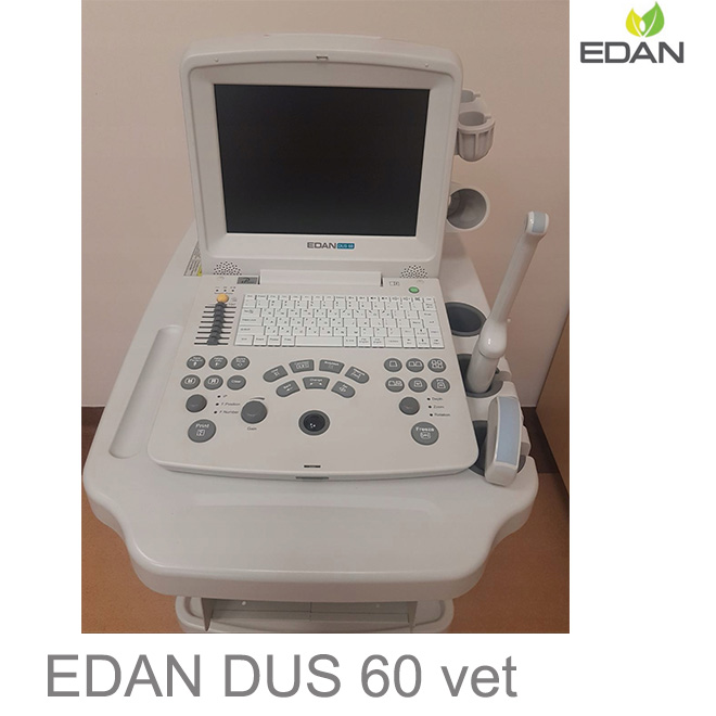
Two standard transducer connectors.
Maximum frequency up to 10 MHz.
12.1" TFT-LCD monitor.
Folding keyboard designed with trackball is easy and convenient for various types of operation.
Two USB ports support storage, transferring, and printing.
256-frame bidirectional cine loop memory.
8-segment TGC control.
IP (Image Processing) Function.
Measurement and calculation software packages.
General:
Gray Scales: 256.
Scanning Angle: Up to 152° (transducer dependent).
Scanning Depth: From 19 mm to 324 mm (transducer dependent).
Functions:
Body Mark: > 130 types.
Transducer auto-detection.
Peripheral Ports: S-video output, Video output, VGA output, two USB ports, Ethernet port, Remote control, and Footswitch port.
Measurement and Calculation:
B-mode: Distance, circumference, area, volume, angle, ratio, %stenosis, histogram.
M-mode: Distance, time, heart rate, slope.
Doppler: Time, heart rate, velocity, acceleration, trace, and RI.
Power Supply: 100 - 240 V (50/60 Hz).
Dimensions: 13" (L) x 8.7" (W) x 12.6" (H) (330 mm x 220 mm x 320 mm).
Net Weight: 15.7 lbs (7.1 kg).


 Price is 8-20% Lower Than Other




![{pr0int $v['title']/}](https://medicalequipment-msl.com/upload/img/20150605/20150605153036394.jpg.jpg)
![{pr0int $v['title']/}](https://medicalequipment-msl.com/upload/img/20180103/201801031738207058.jpg.jpg)
![{pr0int $v['title']/}](https://medicalequipment-msl.com/upload/img/20151112/201511121625043859.jpg.jpg)
![{pr0int $v['title']/}](https://medicalequipment-msl.com/upload/img/20130715/201307151839437991.jpg.jpg)
![{pr0int $v['title']/}](https://medicalequipment-msl.com/upload/img/20130830/201308301133451351.jpg.jpg)
![{pr0int $v['title']/}](https://medicalequipment-msl.com/upload/img/20140311/201403111759353202.jpg.jpg)
![{pr0int $v['title']/}](https://medicalequipment-msl.com/upload/img/20130716/201307161605509836.jpg.jpg)
![{pr0int $v['title']/}](https://medicalequipment-msl.com/upload/img/20130716/201307161021424404.jpg.jpg)
![{pr0int $v['title']/}](https://medicalequipment-msl.com/upload/img/20180228/20180228155012511.jpg.jpg)
![{pr0int $v['title']/}](https://medicalequipment-msl.com/upload/img/20141107/201411071545146803.jpg.jpg)
![{pr0int $v['title']/}](https://medicalequipment-msl.com/upload/img/20151203/201512031810234996.jpg.jpg)
![{pr0int $v['title']/}](https://medicalequipment-msl.com/upload/img/20130716/201307161005073463.jpg.jpg)
![{pr0int $v['title']/}](https://medicalequipment-msl.com/upload/img/20170620/20170620155056520.jpg.jpg)
![{pr0int $v['title']/}](https://medicalequipment-msl.com/upload/img/20180228/201802281539286385.jpg.jpg)
![{pr0int $v['title']/}](https://medicalequipment-msl.com/upload/img/20191105/201911051730429327.jpg.jpg)
![{pr0int $v['title']/}](https://medicalequipment-msl.com/upload/img/20190123/201901231522213839.jpg.jpg)
![{pr0int $v['title']/}](https://medicalequipment-msl.com/upload/img/20190123/201901231706538520.jpg.jpg)
![{pr0int $v['title']/}](https://medicalequipment-msl.com/upload/img/20191105/201911051524074406.jpg.jpg)
![{pr0int $v['title']/}](https://medicalequipment-msl.com/upload/img/20191105/201911051629267630.jpg.jpg)
![{pr0int $v['title']/}](https://medicalequipment-msl.com/upload/img/20191105/201911051716533960.jpg.jpg)
![{pr0int $v['title']/}](https://medicalequipment-msl.com/upload/img/20191105/201911051703061617.jpg.jpg)
![{pr0int $v['title']/}](https://medicalequipment-msl.com/upload/img/20191107/201911071053219520.jpg.jpg)
![{pr0int $v['title']/}](https://medicalequipment-msl.com/upload/img/20191206/201912061030567326.jpg.jpg)
![{pr0int $v['title']/}](https://medicalequipment-msl.com/upload/img/20191226/201912262246159805.jpg.jpg)
![{pr0int $v['title']/}](https://medicalequipment-msl.com/upload/img/20191228/201912282217384903.jpg.jpg)


