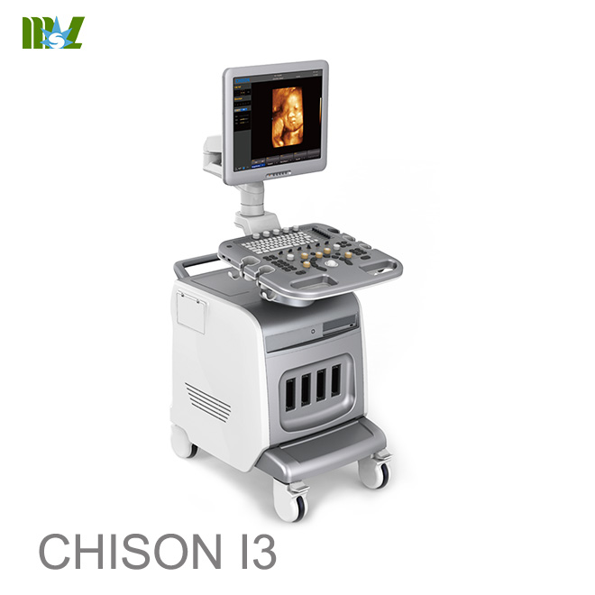
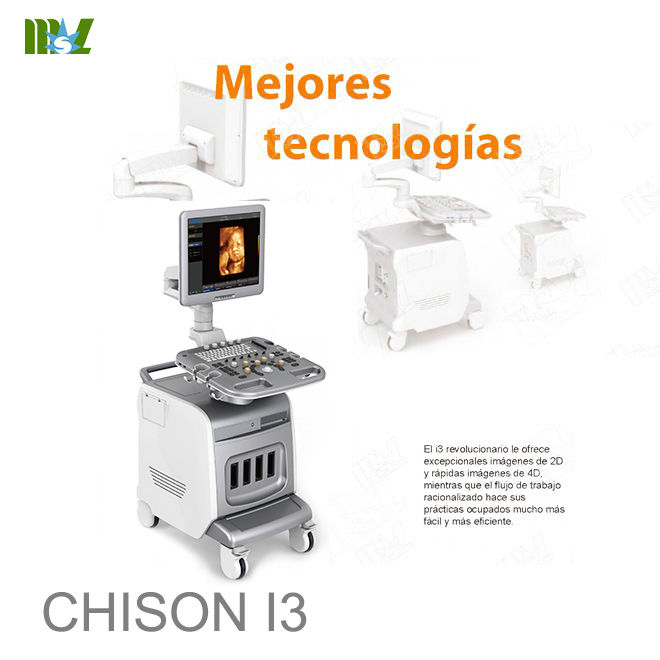
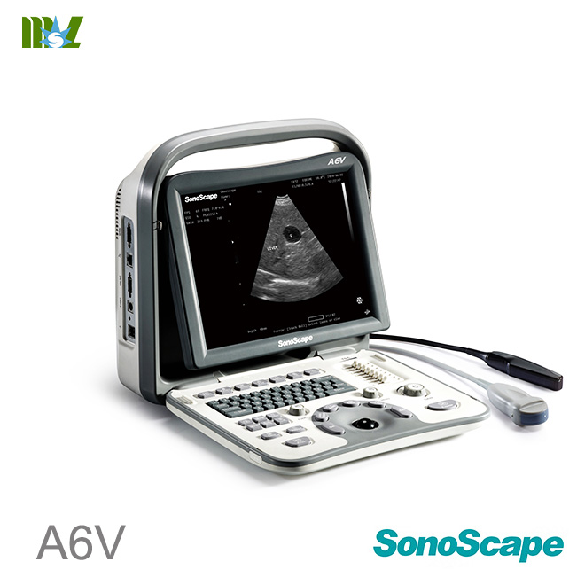
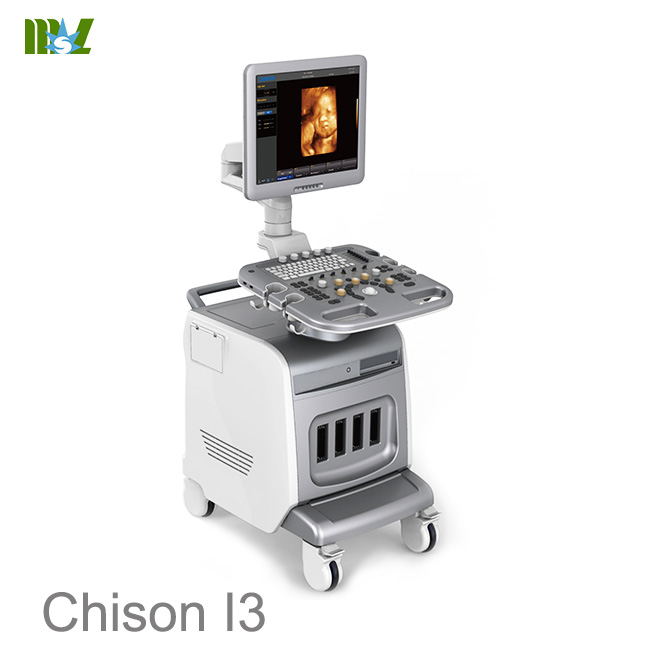
ultrasonido 4d doppler obstetrico : ultrasonido abdominal chison i3
ultrasonido 4d doppler obstetrico chison i3 IMAGING MODES
* B, 2B, 4B, B/M, M
* CFM
* PW Mode
* Power Doppler/Directional PD
* Trapezoidal
* Real-time 4D (Option)
* Chroma B/PW
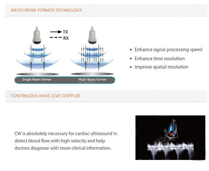
* CW (Option)
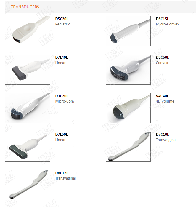
ultrasonido 4d doppler obstetrico chison i3 PROBES
* Convex probe
* Linear probe
* Transvaginal probe
* Phased array probe
* Micro-Convex probe
* 4D Volume probe
ultrasonido 4d doppler obstetrico chison i3 IMAGE PROCESSING TECHNOLOGY
* AIO(Automatic adjust dynamic motion)
* Trapezoidal imaging
* SRA(Speckle noise reduction
* THI
* Compound technology
* i-lmage: image optimization software
ultrasonido 4d doppler obstetrico chison i3 MEASUREMENT & REPORT PACKAGES
* OB&GYN
* Vascular
* Urology
* Small parts
* Cardiac
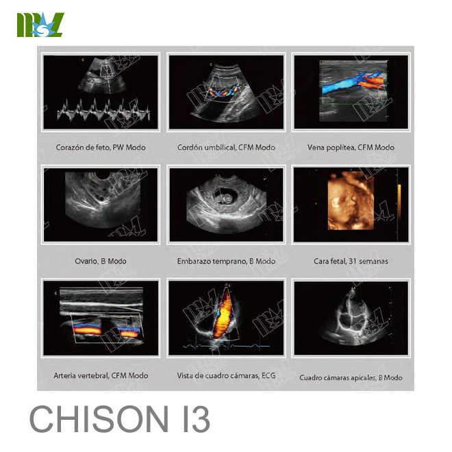
ultrasonido 4d doppler obstetrico chison i3 WIDELY CLINICAL APPLICATIONS
* Abdominal
* * Vascular
* Peripheral vascular
* Small parts
* Prostate
* Breast
* Superficial
* Musculoskeletal
* Gynecological and fertility
* Pediatric general imaging
* Surgical Imaging
* Obstetrical
* Interventional Imaging
* Epicardial Imaging
* Fetal Echo
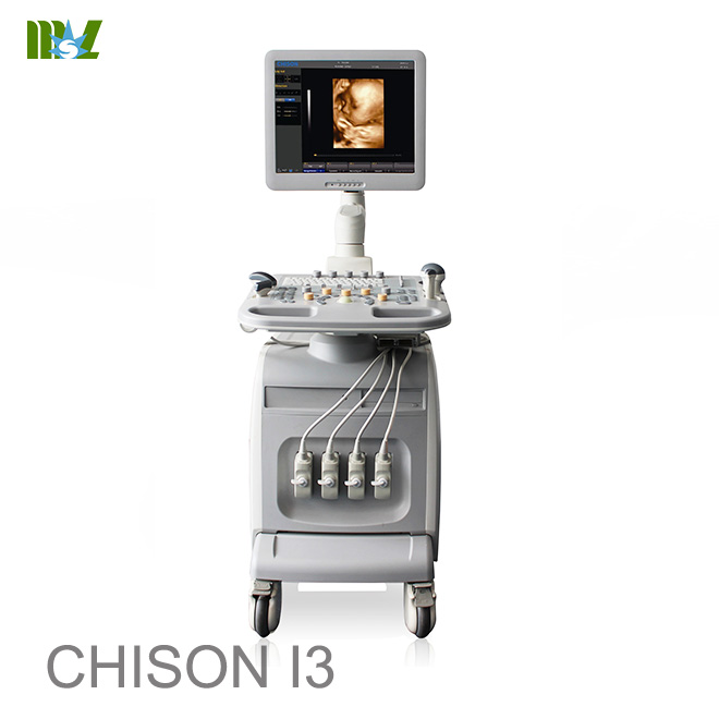
ultrasonido 4d doppler obstetrico chison i3 STANDARD CONFIGURATION
Main unit, 19" LCD,4 probe connectors,
Hard disk, DVD-RW,6 USB flash memory
ultrasonido 4d doppler obstetrico chison i3 OPTIONS
* Convex probe
* Linear probe
* Linear probe (60mm)
* Transvaginal probe
* Transvaginal probe
* Phased array probe
* Micro-Convex probe
* Micro-Convex probe (adult Cardiac)
* Micro-Convex
* CW
* ECG
* 4D package: induding 2.5MHz-5.3MHz 4D volume probe, 4D software and 4D hardware module
* Video printer(SONY UP-X898MD), PC printer(HP Pro 111P1102W & HP Pro 200 M251n & Canon selphy cp910)
* DICOM 3.0
* i-lmage: image optimization software
* Biopsy kit:for convex, linear,TV probe
* Foot-switch
* Super needle
* 2D steer
* Quadplex
* Auto IMT
* Elastography
We reserve the right to make changes to this document without advance notice
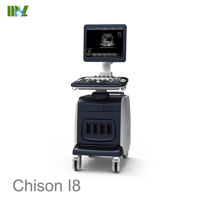
i3
Color Doppler System Datasheet V3.2
We reserve the right to make changes to this document without advance notice
GeneralInformation
Dimensions and Weight
Dimensions of main unit (approx.):
630mm (width)×1020mm (depth) ×1365mm(height)
Net weight of main unit (approx): 125kg(no probe included)
Electrical Power
Power supply voltage: Auto adaptable for AC100-240V
Power supply frequency: 50-60 Hz
Power consumption: 600 VA
Operationpanel
Control panel
Alphanumeric keyboard
8 TGC Slides
Interactive backlit keys
High resolution color LCD
- Diagonal dimension: 19 inch
- Resolution: 1280X1024
- Brightness adjustment
Integrated speaker
- Volume adjustable
SystemOverview
Applications
Abdomen
Gynecology
Obstetrics
Urology
Small Part
We reserve the right to make changes to this document without advance notice
Pediatrics
Vascular
Musculoskeletal
Cardiac
Scanning Method
Electronic convex
Electronic linear
Electronic micro convex
Electronic phased array
Volume convex
TransducerTypes
3.5MHz Convex probe, D3C60L
7.5MHz Linear probe, D7L40L
6.0MHz Transvaginalprobe, D6C12L
3.0MHz Micro convex Probe D3C20L
6.0MHz Micro convex probe D6C15L
3.0 MHz Phased array probe D3P64L
4.5MHz 4D volume probe V4C40L
Image Modes
B mode
M mode
PW (Pulse Wave Doppler)
CW (option)
CFM (ColorFlow Mapping)
CPA (Power Doppler)
DPD (DirectionalPower Doppler)
4D (option)
ECG (option)
Trapezoidal imaging (onlyfor linear probe)
Display Mode
We reserve the right to make changes to this document without advance notice
Quad/dual display (for B, CFM, CPA)
Duplex mode: B+CFM, B+PW, B+CPA,B+DPD,B/M
Display Annotation
Hospital name
Date/Time
Patient Name and Patient ID
System status (real-time or frozen)
Gray/Color bar
Cine guide
Scanning direction
Measurement summary window
Measurement results window
Probe type
Frequency
Application name
Menu indication
Trackball functions indication
Imaging parameters displayed on the screen
Standard Configuration
High resolution 19 Inch LCD display
4 activeprobe ports
Pulse Wave Doppler
Color Doppler Flow Imaging
Power Doppler Flow Imaging
Directional Power Doppler Flow Imaging
≥250Gintegrated hard disk
USB ports: 6(2 at the control panel, 4 at the rear panel)
Ethernet port
S-video out port
VGA port
General measurement package
Clinical measurement package
We reserve the right to make changes to this document without advance notice
Multi-language screen display
EasyView: image archivesystem
Patient information management system
Building reporting system
AIO (Automatic Image Optimization)
Intelligent Zoom
Speckle Reduction Algorithm (SRA)
i-Image TM software package
Software Options
DICOM 3.0
4D software package
CW
ECG
Hardware Option
3.5MHz Convex probe, D3C60L
7.5MHz Linear probe, D7L40L
6.0MHz Transvaginalprobe, D6C12L
3.0MHz Micro convex Probe D3C20L
6.0MHz Micro convex probe D6C15L
3.0MHz Phased array probe D3P64L
4.5MHz 4D volume probe, V4C40L and4D module
Footswitch
ECG Cable
Peripherals
Video printer: SONY UP897MD
PC printer :
- HP Laser Jet 1020
- HP Laser Jet CP2055d
We reserve the right to make changes to this document without advance notice
Imaging Processing and Presentation
B Mode
Acoustic power
Gain
TGC
Depth
Freq.
Frame rate
Focus number
Focus position
Scan width
Line density
Dynamic
Persistence
Noise reject
Smooth
Edge enhance
i-Image TM
SRA
Compound
2D Map
Chroma
Gamma
Screenbrightness
Image rotate
Zoom
M Mode
Color Map
Sweep speed
Layout
We reserve the right to make changes to this document without advance notice
Color Mode
Gain
Frame rate
Steer
PRF
Wall filter
Color Map
Flow
Color Invert
Density
Persistence
Baseline
Color mode:Velocity, Variance
BloodEffection
Scale
CPA/DPD Mode
Gain
Frame rate
Steer
PRF
Wall filter
Color Map
Flow
Density
Persistence
Wall Thre.
PW Mode
Gain
PRF
Scale
Invert
Wall Filter
We reserve the right to make changes to this document without advance notice
Audio
Speed
Baseline
DA
SV
Color Map
2D Map
CW Mode
Gain
PRF
Scale
Invert
Wall Filter
Audio
Color Map
Speed
Baseline
2D Map
Dynamic
Spectrum Enhance
DA
Storage
≥250GB integrated hard drive
DVD R/W driver
USB ports
Still images storage format: IMAG
Still images export format: BMP, JPG, DCM,PNG,TIFF
Cine loops storage format: CINE
Cine loops export format:AVI
Fast storage setting:3s,5s,10s,customize time ,manual
We reserve the right to make changes to this document without advance notice
EasyView
Image review Layout:1×1,2×2
Image management
- Delete selected image
- Export selected image
- Send selected image to demo
- Print selected image by PC printer
- Print selected image by DICOM printer
- Send selected image by DICOM
- Selected all
- Selected none
Exam Review
SearchExam
Exam review:patient view, study view
Exam management
- Delete selected exam
- Export selected exam
- Backup selected exam
- Recover from the backup exam
- Selected all
- Expand all
- Collapse all
- Edit selected Exam
- Review selected Exam
- Continue selected Exam
Measurement & Calculation
General Measurement Package
- Software packages for various specific clinical use
- Comprehensive analysis methods
- Clinical analysis reports
We reserve the right to make changes to this document without advance notice
General measurement package
B modeNormal measurement
Distance
Length_Area(Ellipse)
Length_Area(Trace)
Volume(1 Distance)
Volume(2 Distance)
Volume(3 Distance)
Volume(1 Ellipse)
Volume(2 Ellipse)
Volume(1 Distance 1 Ellipse)
Ratio
Angle
M mode Normal measurement
Mdistance
Mtime
Velocity
Heart_Rate
PW mode Normal measurement
Velocity
Distance
Peak
Auto Trace
Manual Trace
HR
Flow Volume
StD%
StA%
Area
Clinical Analysis Packages
OB
OB –B measure
Distance
Fetal Biometry:GS,CRL,YS,BPD,OFD,HC_Ellipse,APD,TAD,AC(Ellipse),
We reserve the right to make changes to this document without advance notice
FTA,FL,SL,APTD,TTD,ThC
Fetal Long Bones:Humerus,ULNA,Tibia,RAD,FIB,CLAV
Fetal Cranium:CER,CM,NF,NT,OOD,IOD,NB,Lvent,HW
OBOthers:LtKid,RtKid,LtRenalAP,RtRenalAP,LVWrHEM,MAD
AFI:AFI_1,AFI_2,AFI_3,AFI_4
FBP:AF
Ductus Venosus:StA%,StD%,Vessel Area,Vessel Dis
StA%:A Out,A In
StD%:D Out,D In
CX_L
Aorta:StA%,StD%,Veslumen_D,Veslntimal_D,VesOutside_D,Veslntimal_A,
Veslumen_A
StA%:A Out,A In
StD%:D Out,D In
Descending Aorta:StA%,StD%,Veslumen_D,Veslntimal_D,VesOutside_D,
Veslntimal_A,Veslumen_A
StA%:A Out,A In
StD%:D Out,D In
MCA:StA%,StD%, Veslumen_D,Veslntimal_D,VesOutside_D,Veslntimal_A,
Veslumen_A
StA%:A Out,A In
StD%:D Out,D In
UmbA:StA%,StD%,Veslumen_D,Veslntimal_D,VesOutside_D,Veslntimal_A,
Veslumen_A
StA%:A Out,A In
StD%:D Out,D In
Uterine Artery:Uterine Artery (Rt), Uterine Artery (Lt)
Uterine Artery (Rt): StA%,StD%, Veslumen_D,Veslntimal_D,VesOutside_D,
Veslntimal_A,Veslumen_A
Uterine Artery (Lt): StA%,StD%, Veslumen_D,Veslntimal_D,VesOutside_D,
Veslntimal_A,Veslumen_A
StA%:A Out,A In
StD%:D Out,D In
Pulmonary Artery:StA%,StD%,Veslumen_D,Veslntimal_D,VesOutside_D,
Veslntimal_A,Veslumen_A
StA%:A Out,A In
We reserve the right to make changes to this document without advance notice
StD%:D Out,D In
Fetal Select
OB–D measure
Umb A
Aorta
Descending Aorta
Uterine Artery (Lt)
Uterine Artery (Rt)
Pulmonary Artery
MCA
FHR
OB –M measure
Mdistance
Mtime
Velocity
Heart_Rate
GYN
GYN –B measure
Distance
UT:UT_L,CX_L,UT_W,UT_H
Cervix Vol.:Length,Height,Width
ENDO
Right_OV_Volume:Length,Height,Width
Left_OV_Volume:Length,Height,Width
Right FO_D:Length,Width
Left FO_D:Length,Width
Uterine Artery:Uterine Artery(Rt),Uterine Artery(Lt),
UterineArtery(Rt):StA%,StD%,Vessel Area,Vessel Dis
UterineArtery(Lt):StA%,StD%,Vessel Area,VesselDis
StA%:A Out,A In
StD%:D Out,D In
GYN –Dmeasure
Umb A:Umb A(Rt),Umb A(Lt)
MCA:MCA(Rt),MCA(Lt)
Rt UterineA
Lt Uterine A
FetalAO:Fetal AO(Rt),FetalAO(Lt)
FHR
GYN –M measure
We reserve the right to make changes to this document without advance notice
Mdistance
Mtime
Velocity
Heart_Rate
Pediatrics
HIP
URO
Distance
Residual Vol.
Prostate Vol.
Left Kidney
Right Kidney
T-Zone Vol.
Bladder Vol.
StA%
StD%
Vessel Area
Vessel Dis
Cardiac
Cardiac-B measure
Distance
SinglePlane:
BiPlane
Bullet_Volume
Modi_Simpson
Teichholz
Cube
LV/RV
AO/LV
LVOT
MV
AV
Cardiac-D measure
We reserve the right to make changes to this document without advance notice
LVOT
AV
MV
TV
PV
Pul.Vein
HR
Cardiac-M measure
Distance
Heart_Rate
Ejection_Time
LV
LVSHORT
AV
AVSHORT
MV
AV
AO/LV
LVOT
TV
PulV
Vessel
Prox CCA
Mid CCA
Distal CCA
Prox ICA
Mid ICA
Distal ICA
ECA
Vertebral A
INT IIL
EXT IL
ILIAC
CFA
ProFun
LTCIR
SFA
We reserve the right to make changes to this document without advance notice
Pop A
ATA
PTA
PERON
DRPED
Abdomen
CBD
GB Wall
Liver Length
Bladder
Prox Aorta
Mid Aorta
Distal Aorta
Spleen
Renal Vol.
Lliac
Carotid
Subclavian A
Prox CCA
Mid CCA
Distal CCA
Bulb
Prox ICA
Mid ICA
Distal ICA
ECA
Vertebral A
General Measurement
Flow Volume
Small parts
General Measurement
Ratio
We reserve the right to make changes to this document without advance notice
Angle
System Setup
Byusing system Setup, userscould
Customize hospital information
Customize language
Customize fast storage time
Customize colormap
Assign functions to “PRINT” button on control panel and foot switch
Customize comment library
Customize report
User Define Functions
Byuser-define function, users could customize user-define preset, including
- Applications name, Presets name, User defined name
- Applications exam type
- Imaging parameters
Multi-language Display Interface
English
Chinese
French
Spanish
Russian
Polish
Portuguese
Operation System
Windows XP Embedded
We reserve the right to make changes to this document without advance notice
Transducers
Transducer Selection
Name Array type Center
frequency
D3C60L Convex 3.5MHz
D7L40L Linear 7.5MHz
D6C12L Micro-convex 6.0MHz
V4C40L Convex 4.5MHz
D3C20L Micro-convex 3.0MHz
D6C15L Micro-convex 6.0MHz
D3P64L Phased Array 3.0MHz
Inputs & Outputs
S-video: 1
Video out: 1
VGA: 2
USB port: 6
Ethernet: 1
Remote control: 1
Footswitch port: 2
System power in: 1
Ground pole: 1
Power button: 1
Operating Conditions
Ambient temperature: 10°C to 40°C
Relative humidity: 30% to 75% (no condensation)
Atmospheric pressure: 700 hPa to 1060 hPa
Storage Conditions
Ambient temperature: -5°C to 40°C
Relative humidity: ≤ 80% (no condensation)
We reserve the right to make changes to this document without advance notice
Atmospheric pressure: 700 hPa to 1060 hPa
Quality Standards
ISO 10993 Biological evaluation of medical devices
IEC 60601-1 Electrical medical equipment
IEC 60601-1-1 Electrical medical equipment
IEC 60601-1-2 Electromagnetic compatibility
IEC 60601-1-4 Programmable medical systems
IEC 60601-2-37 Particular requirements for the safetyof ultrasonic medical
diagnostic and monitoring equipment.
Not all features or specifications described inthis document may beavailable in all probes
and/or modes.
CHISON Medical ImagingCo., Ltd. reserves the right to make changes in specifications
and features shown here in, or discontinues the product atany time without notice or
obligation. For the most current information,Contact CHISON Representative.
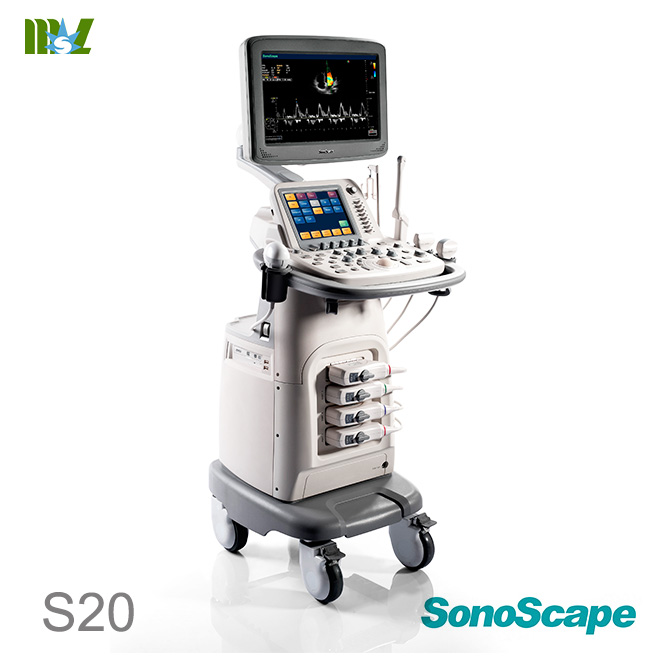
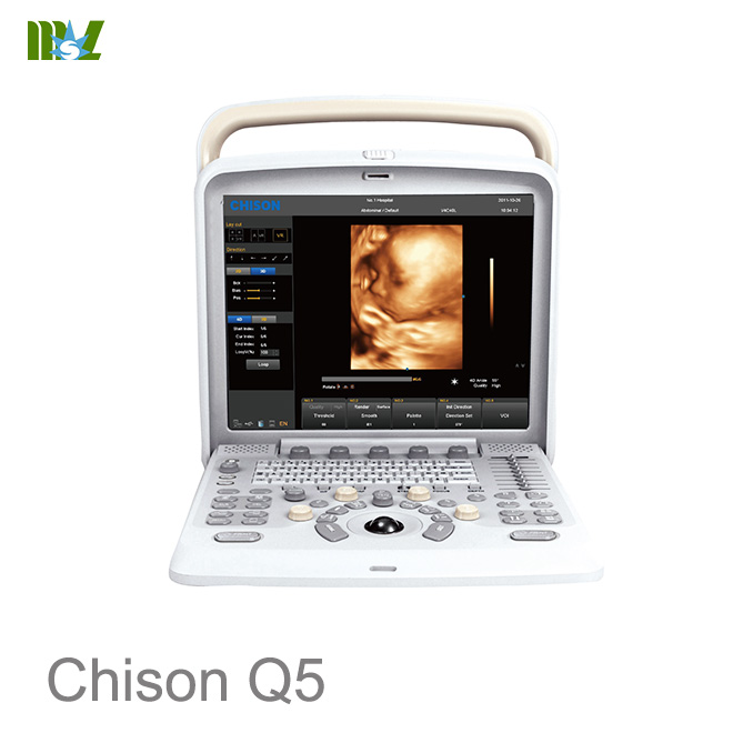
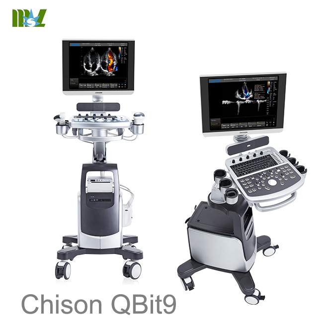
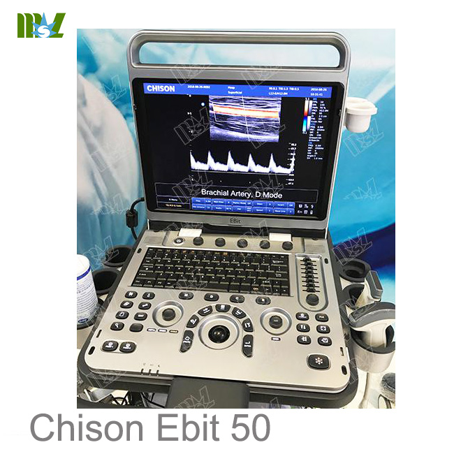
Hot sale Sonoscape ultrasound | Chison ultrasound price list
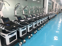
MSL TEAM picture


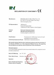
MSL Certificate
MSL Medical cooperate with DHL,FEDEX,UPS,EMS,TNT,etc.International shipping company,make your goods arrive destination safely and quickly.


 Price is 8-20% Lower Than Other