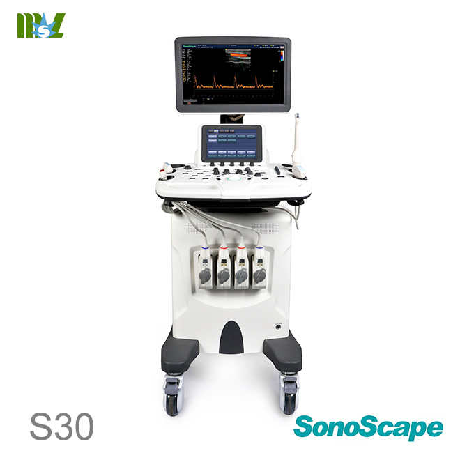
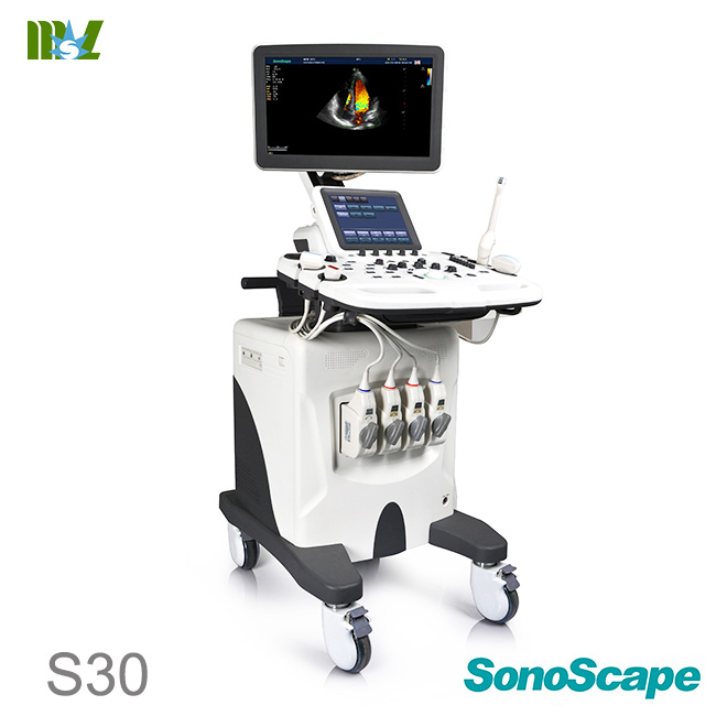
ecografie endovaginala SonoScape S30 Standard Configurations
Standard Hardware include:
S30 main unit
19" High Resolution LED color monitor
10.4" High Resolution Touch Screen
Four transducer connectors
One CW transducer connector
DVD-RW/ USB 2.0/ Hard Disk 500 G
ECG Module
Standard Software include:
Imaging modes: B/ 2B/ 4B/ M/ THI/ CFM/ PDI/ DirPDI/ PW/ HPRF/ CW
LGC: Lateral gain compensation
Pulse Inversion Harmonic
Trapezoidal Imaging
Compound Imaging
μ-Scan: 2D speckle reduction technology
Multi-beam technology
Auto NT
Freehand 3D Imaging
S-Depth
S-Live
Advanced cardiovascular kit: TDI/ Color M/ IMT/ Steer M/ Auto EF
Stress Echo
VIS-Needle
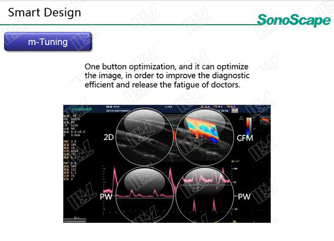
M-Tuning: one button image optimization
DICOM 3.0: Store/C-Store/Worklist/MPPS/ Print/ Q/R
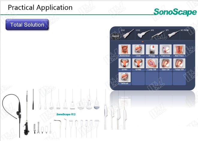
Stardard Configured Transducers:
192 elements linear array L742 (Vascular, Small parts, MSK etc.), 3.5-16MHz/38mm
64 elements phased array 2P1 (Cardiac, Transcranial), 1-6MHz
128 elements convex array 3C-A (Abdominal, Obstetrics, Gynecology), 1.0-7.0MHz/ R50mm
192 elements endocavity 6V3 (Gynecology, Obstetrics, Urology), 3-15MHz/ R10mm
ecografie endovaginala SonoScape S30 video
ecografie endovaginala SonoScape S30 Optional Configurations
Static 3D
4D
Contrast Imging
C-xlasto: Elastography Imaging
2D Panoramic Imaging
Color Panoramic Imaging
ecografie endovaginala SonoScape S30 General Specification
S22 digital color Doppler ultrasound system adopts the advanced ultrasonic Doppler technologies, including the Full Digital Super-wide Band Beam Former, Digital Dynamic Focusing, Variable Aperture and Dynamic Tracing, Wide Band Dynamic Range, Multi-beam Processing, etc. The ultrasound diagnostic software in ergonomic design can be customized and easily performed by users. Based on the computer technology and Linux operating system, this ultrasound system is reliable and stable. System maintenance and upgrade can be completed by updating software to achieve product improvements and advanced technology. Complied with the international standards and regulations, this ultrasound system is safe and effective.
ecografie endovaginala SonoScape S30 Advanced Technologies
Digital front-end technology
Multi-beam forming technology
Compound imaging
µ-scan image processing
Tissue harmonic imaging
Phase-inversion harmonic imaging
High pulse repetition frequency
Panoramic imaging
4D imaging
Exam-type icons
Touch screen
Elastography Imaging
Contrast imaging
ecografie endovaginala SonoScape S30 Standard Configurations
Touch screen
DVD
ECG Module
µ-scan
5-band adjustable frequency in B mode
LGC (2-band)
Tissue characteristic index
THI mode
PIH mode
Dual beams
Image rotation function
Compound imaging
Trapezoidal imaging
Color mode
PDI mode
DPDI mode
PW mode
HPRF Support
Simult mode
FreeHand 3D
Steer M mode
Biopsy enhanced
Basic measurement package
Obstetrics measurement package
Gynecology measurement package
Cardiology measurement package
Abdomen measurement package
Vascular measurement package
Urology measurement package
Small parts measurement package
Pediatrics measurement package
Myocardial performance index
PW auto trace
IMT measurement
DICOM transmission
Digital Color Doppler Ultrasound System Specifications
4 /29
901-01657-A05
DICOM worklist
DICOM MPPS
DICOM C-store
DICOM Q/R
4. Optional Functions
4D imaging
Bi-plane probes available in the real-time dual
mode
Color M mode
TDI mode
CW mode
Stress Echo
Panoramic imaging in 2D mode
Panoramic imaging in color flow mode
Elastography Imaging
5. Optional Accessories
Biopsy guide brackets
Color ink jet printer
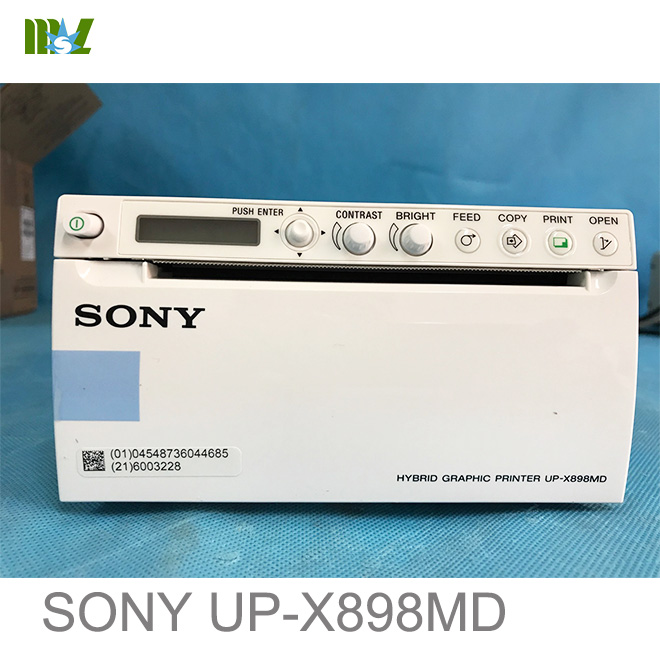
B/W video printer
Color video printer
Transducer cable hanger
Footswitch
6. Scan Methods
Electronic curved array
Electronic linear array
Electronic phased array
Mechanical sector scan
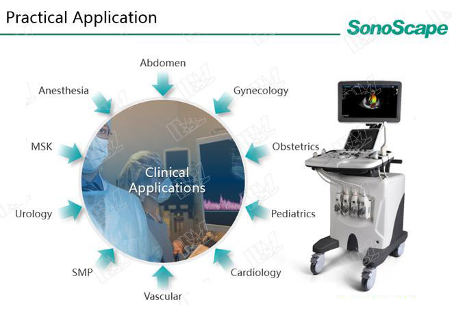
7. Applications
Abdomen
Vascular
Cardiology
Gyn/OB
Urology
Musculoskeletal
Interventional ultrasound
Small parts
Anesthesiology
Pediatrics
Orthopedics
Cephalic
8. Imaging Modes
B mode
M mode
THI mode
CFM mode
PDI mode
TDI mode
PW mode
CW mode
HPRF
3D/4D mode
Color M mode
Steer M mode
Elastography Imaging
9. Display Formats
B+B
4B
B + Color
B + Color (Dual real time)
B + PDI
Digital Color Doppler Ultrasound System Specifications
5 /29
901-01657-A05
B + TDI
B + M
B + Color + M
B + PDI + M
B + TDI + M
B + PW
B + Color + PW
B + PDI + PW
B + TDI + PW
B + CW
B + Color + CW
B + PDI + CW
Panoramic imaging
Trapezoidal imaging
10. System Settings
General Settings
Hospital Name
Language
Freeze Response: Cine, Comment,
Measure, Body Mark, Arrow, Distance
Trackball Sensitivity: 1, 2, 3, 4, 5, 6
Date/Time Setting
Monitor Type: TV-NTSC, TV-PAL,
VGA(4:3), VGA(16:9)
Caps Lock: On, Off
Clear Annot On Unfreeze: On, Off
Guide Line Type: Gun, Needle
Focal Auto: On, Off
Display Settings
Color of ROI: Green, Yellow, Orange,
Cyan
Display Format: V1/2, V1/3, V2/3, H1/2,
H1/4, O1/4
Screen Save: On, Off
Screen Saver Delay: 0-99minutes
Annot Font Size: Large, Medium, Small
Storage Settings
Clip Format: WMV, AVI
Still Format: JPG, BMP, TIF
Store Frame Amount: 100-1000 frames
Print&Store Region: Full Screen,
Image&Result Area, US Image
Image Share Service: On, off
Store To UsbDisk
Key Configuration
Save Image: Print, Send to Usb, DICOM
Send, DICOM Print
Save Cine: Send to Usb, DICOM Send
P1 shortcut key: save image, cine,
volume data
P1 shortcut key: automatically response
to print, send to Usb, DICOM Send,
DICOM Print
P2 shortcut key: save image, cine,
volume data
P2 shortcut key: automatically response
to print, send to Usb, DICOM Send,
DICOM Print
Peripheral Device Settings
Local Network Settings
DHCP or Static IP
Static IP: IP Address, NetMask,
Default Gateway, DICOM AE Title,
Mac Address
Advance: Speed (10M, 100M, 1000M,
Unknown); Duplex (Semi Duplex, Full
Duplex, Unknown)
Digital Color Doppler Ultrasound System Specifications
6 /29
901-01657-A05
Ping IP Address
Printer Settings
Printer Driver
Video Invert
Default Printer
Net Printer IP
Comment Settings
Classified by specific exams
Edit
Delete
Arrange
Bodymark Settings
Classified by specific exams
Up to 135
Measurement Settings
General Measurement Items
BSA setting: Eastern, Western
Cross Cursor Size: Small, Medium,
Large
Measure Line Size: Small, Medium,
Large
Distance Dash Line Display: On, Off
Velocity Cross Line Display: On, Off
Ellipse Cross Line Display: On, Off
Line ID Display: On, Off
Keep Result Window: : On, Off
Freeze on Measure: On, Off
10 shortcut keys are available for
OB/GYN exams
Calculation Formula
EFW
Age by EFW
EFW GP/SD
CUA
BPD
OFD
HC
AC
FL
HL
Cereb
CRL
GS
Clav.
RAD
Ulna
TIB
FIB
BOD
Touch-Screen Settings
Select Probe (Curved array, linear array,
phased array, pencil probe)
Select Mode (B/M/CFM/PDI/PW mode for
curved or linear array probe;
B/M/CFM/TDI/PDI/PW/CW mode for
phased array probe; PW/CW mode for
pencil probe)
Select Status (real-time or frozen mode)
Arrange items on the touch screen
Load default
Report Settings
Logo
Operation Logo (insert logo from the USB
drive)
Title/Font
Title1 (font size can be set to 14, 16, 18,
20, 22, 24, 26)
Title2 (font size can be set to 14, 16, 18,
20, 22, 24, 26)
Digital Color Doppler Ultrasound System Specifications
7 /29
901-01657-A05
Title3 (font size can be set to 14, 16, 18,
20, 22, 24, 26)
Context Title (font size can be set to 12,
13, 14, 15, 16)
Subtitle (font size can be set to 14, 16, 18,
20, 22, 24, 26)
Background Color
Text Color
Display Items For Report
Patient Information
Exam Information
Measurements (optional)
Image (optional)
Graphs (optional)
Comment
Preview
DICOM
Store
Service List
Remote Host Name, IP Address,
DICOM AE Title, Port Number,
Connect Timeout (sec), Repeat Count,
Dimse Timeout (sec), Acse Timeout
(sec), Send After Every Image Stored,
Send At End of Exam
Commitment
Service List
Remote Host Name, IP Address,
DICOM AE Title, Port Number,
Connect Timeout (sec), Repeat Count,
Dimse Timeout (sec), Acse Timeout
(sec), Synchronously Receive
N-EVENT-REPORT Message
Worklist
Service List
Remote Host Name, IP Address,
DICOM AE Title, Port Number,
Connect Timeout (sec), Repeat Count,
Dimse Timeout (sec), Acse Timeout
(sec), Max. Result
MPPS
Service List
Remote Host Name, IP Address,
DICOM AE Title, Port Number,
Connect Timeout(sec), Repeat Count,
Dimse Timeout(sec), Acse
Timeout(sec)
Print
Service List
Remote Host Name, IP Address,
DICOM AE Title, Port Number, Format,
Trim, Priority, Border, Medium, Empty
Image, Orientation, Color, Film Size,
Min Density, Film Destination, Max
Density, Magnification, Configure
Information, Copies, Film Session
Label, Smoothing Type
QR List
Service List
Remote Host Name, IP Address,
DICOM AE Title, Port Number,
Connect Timeout (sec), Repeat Count,
Dimse Timeout (sec), Acse Timeout
(sec), Max. Result
Load Default
Load
New
Export/Import
System Information
11. System Parameters
Frame rate: 935 fps/s (probe dependent)
Digital Color Doppler Ultrasound System Specifications
8 /29
901-01657-A05
Grayscale Level: 256
Transducer Elements: 256
12. B Mode
Gain: 1-255 adjustable
Scan Depth: 42.9cm
Image Zoom, Showing zoom ratio (0.8-10
times)
TGC: 8 levels slider controls
Image Inversion: Left and Right, Up and Down
Panoramic imaging: achievable
Compound imaging: Off, 1, 2 adjustable
Focus: Up to 12, Focus span adjustable (probe
dependent)
Frequency: 5 bands adjustable
Chroma: 13 types selectable
Adaptive image fusion: 15 types selectable
μ-Scan: 0, 2, 3, 7, 11 adjustable
Line Density: 3 levels adjustable
(High/Medium/Low)
Persistence: 0-95 selectable
Biopsy Guide Function: On/Off
Biopsy lines angle adjustable
Biopsy lines offset adjustable
Dynamic Range: 20-280 (Probe dependent)
Grayscale Curve: 7 selectable
Imaging width and position: adjustable
Power: 1-100 adjustable, one step each
Tissue Acoustic:1400-1700
LGC: gain adjustable on the left/right part
Trapezoid Imaging: On/Off (Linear array probe)
B steer Mode (Linear array probe)
m-tuning
13. Color/TDI
Gain: 0-255
Frame Rate: 50 frames/sec
Size and position of color ROI: adjustable
Auto Focus (focus number:1)
Inversion: Up/Down, Left/Right
Flow Invert: On/Off
Frequency Range: 5 steps, adjustable
Wall Filter: 25-750Hz (probe dependent)
PRF: 0.5-12kHz (Probe dependent)
Line Density: 4 kinds
(low/medium/high/max-high)
Color/Direction energy: 10 kinds selectable for
color Doppler, 4 kinds selectable for tissue
Doppler
Color baseline adjustment: ±15 steps
Persistence: 0-80 (Probe dependent)
B Reject: 0-255 adjustable
Linear deflection angel: 0, ±16, ±20 adjustable
Color Flow: available in the frozen mode
m-tuning
14. M Mode
Steer M: 3 sample lines, Display frame rate
Video Inversion (On/Off)
Chroma: 5 types
Display Format: H1/2, H1/4, V1/3, V1/2, V2/3,
O1/4
Scan Speed: 6 levels adjustable
M Processing: Switch between average and
peak values
Power: 30-100 adjustable
15. PW/CW Mode
Doppler Methods
Digital Color Doppler Ultrasound System Specifications
9 /29
901-01657-A05
PW (pulsed wave) Doppler
CW (continuous wave) Doppler
Triplex: On/Off
Sample Volume and Position for PW Doppler:
0.7-21mm adjustable
Video Inversion: On/Off
Spectrum Inversion: Achievable
θ Angle Correction: 0°, 60°, -60° adjustable
Spectral Real-time Trace: Achievable
Baseline Shift:17 steps selectable
Frequency Range: 5 steps
Wall Filter: 25-750 adjustable
PRF: 1-16kHz (PW) (probe dependent)
PRF: 1-48KHz (CW) (probe dependent)
Max Velocity Range:
0.0004-18 m/s (PW) (probe dependent)
0.0013-63 m/s (CW) (probe dependent)
Scan Speed: 4 kinds selectable
Doppler Chroma: 5 kinds selectable
One-key Auto Optimization
Auto Adjusting Baseline
Auto Adjusting PRF
Auto Correcting Angle
Dynamic Range: 10 kinds selectable
Display Format:H1/2, H1/4, V1/3, V1/2, V2/3,
O1/4
Deflection Angle: 0, ±16, ±20, 5 levels
adjustable
16. 3D Mode
Display Mode:
Dual Planes
Quad Planes
3D Full Display
Adjust Slice
VOI: On/Off
ROI Size: adjustable
ROI sample line curvature: adjustable
Restore: Volume data and ROI sample line
selectable
Scan Mode: Lin, Sec
Z Scale: 0.3-2.0 adjustable (range reduced for
few slices)
Z Axis Angle: 10°-170° adjustable
Orientation: 0°, 90°, 180° and 270°, selectable
Images: A, B, C, 3D image
Render mode: Surface, Skeleton, X-Ray
Clip Plane: On/Off
X Rotation
Y Rotation
Z Rotation
Horizontal Movement: Left/Right
Vertical Movement: Up/Down
Free rotation by trackball operation
Zoom In/Out: adjustable
Transparency: 0-100 adjustable
Contrast: 0-100 adjustable
Brightness: 0-100 adjustable
Smoothness: 0-30 adjustable
Crop: trace (in/out), box (in/out), eraser
(small/big)
Restore the review, undo the last cut
Undo Cut
Auto Rotate: 0°, 45°, 90°, 180° , 270°and 360°,
selectable
View: Top, Bottom, Left, Right, Front, Back
3D Chroma: 0-12 adjustable
B Chroma: 1-13 adjustable
Digital Color Doppler Ultrasound System Specifications
10 /29
901-01657-A05
Reference image: A, B, C
Clip plane: AB, AC, BC, ABC
M-slice: A, B, C
Slice number: 3*3, 4*4, 5*5
Slice Spacing: 0.5-2.0, adjustable
Measurement: Distance, Area, Volume
Store: 3D Image, Volume data
Print
Presets customization
Focal position adjustable
Sweep angle: 5°-75° adjustable
Image Quality: Low/Medium/High
17. 4D Mode
Display Mode:
Dual Planes
Quad Planes
3D Full Display
4D Full Display
ROI sample line curvature: adjustable
ROI Size and Position: adjustable
Focal position: adjustable
Sweep angle: 5°-75° adjustable
Image Quality: Low/Medium/High
Stability: On/Off
Adjust Slice
VOI: On/Off
ROI Size: adjustable
ROI sample line curvature: adjustable
Restore: Volume data and ROI sample line
selectable
Orientation: 0°, 90°, 180° and 270°, selectable
Images: A, B, C, 3D image
Render mode: Surface, Skeleton, X-Ray
Clip Plane: On/Off
X Rotation
Y Rotation
Z Rotation
Horizontal Movement: Left/Right
Vertical Movement: Up/Down
Free rotation by trackball operation
Zoom In/Out
Transparency: 0-100 adjustable
Contrast: 0-100 adjustable
Brightness: 0-100 adjustable
Smoothness: 0-30 adjustable
Crop: trace (in/out), box (in/out), eraser
(small/big)
Restore the review, undo the last cut
Undo Cut
Cine: volume dependant, Max. 0-499 adjustable
Auto Rotation: 0°, 45°, 90°,180°, 270°, 360°
View: Top, Bottom, Left, Right, Front, Back
3D Chroma: 0-12 adjustable
B Chroma: 1-13 adjustable
Reference image: A, B, C
Clip plane: AB, AC, BC, ABC
M-slice: A, B, C
Slice number: 3*3, 4*4, 5*5
Slice Spacing: 0.5-2.0, adjustable
Measurement: Distance, Area, Volume
Store: 3D Image, 3D cine, Volume data
Presets customization
Print
Returnable inactivated mode
18. Physiological Signal Display
ECG Pulse wave
Digital Color Doppler Ultrasound System Specifications
11 /29
901-01657-A05
ECG Lead-three lead system
ECG Gain: adjustable
ECG Position: adjustable
ECG Invert: On/Off
R-Trigger: On/Off
Trigger Delay: adjustable
Frame Count: adjustable
19. Integrated Data Management System
Hard Disk Memory Capacity: 500G
USB Interface: 5 (including a engineering
interface)
20. Image Storage and Playback
Cine playback: up to 1000 frames in B mode
Static and Dynamic image storage in the
real-time single or dual mode
Freely view stored data on PC
Clipboard function (in frozen B mode)
Doppler cine playback: speed is adjustable,
sound can be played back
21. DICOM Network Communication
Storage: Directly transmits images with patient
information to a DICOM file server
Print: Images can be printed directly using a
DICOM compatible printer
DICOM Storage Commitment
DICOM Worklist
DICOM MPPS
DICOM Q/R
Medical digital images and communication
DICOM 3.0 interface
22. Preset Function
Users can customize the presets based on
different probe and diagnostic part to optimize
imaging parameters and adjustment
combination.
Users can arrange the presets.
Users can import or export presets.
23. Patient Data Management
Patient Registration: Name, ID, Gender, Date of
Birth, Height, Weight, LMP, EDD and GA.
Patient data and reports are archived by patient
exams
Reports and images can be previewed
Preview size can be set to 1*1,2*2,3*3
File can be previewed, deleted, used for DICOM
Send and DICOM Print
Files can be exported to the USB drive or DVD.
BMP, JPG, TIF, AVI, WMV are selectable for
export format
24. Annotation and Body Mark Setting
Body mark can be classified by specific exams
Annotation can be selected in the library.
25. Physical Specification
L×W×H (mm): 685mm×520mm×1311mm
Weight: approx. 56kg
Monitor: 18.5’’ Widescreen, anti-flickering,LED
backlight,,can be vertically or horizontally
swiveled
4-idential probe connectors, 1 pencil probe
connector
5 probe holders
Digital Color Doppler Ultrasound System Specifications
12 /29
901-01657-A05
26. Safety Standard
Comply with IEC60601-1, Class I,BF international
standard
27. Environmental Requirements
Operation Environment
Temperature: +10℃ - +40℃
Relative Humidity: 30% - 75% (Non
condensing)
Atmospheric pressure: 700 hPa -1060hPa
Transportation and Storage Environment
Temperature: -20℃ - +55℃
Relative humidity: 20%- 90% (non
condensing)
Atmospheric Pressure: 700 hPa- 1060hPa
Power Supply
100-240V~, 2.7-1.1A
Frequency: 50/60Hz
28. Optional Transducers
Phased Array Probe
2P1
Frequency range: 1.0-6.0MHz
Scan sector: 88°
5P1
Frequency range: 3.0-9.0MHz
Scan sector: 87°
Linear Probe
L752 (4.0-16.0MHz)
L741 (4.0-16.0MHz)
L742 (4.0-16.0MHz)
10I2 (4.0-16.0MHz)
LAP7 (3.0-15.0MHz)
Convex Probe
3C-A (1.0-7.0MHz)
C353 (2.0-6.8MHz)
C322 (2.0-6.8MHz)
C344 (2.0-6.8MHz)
6V3 (3.0-15.0MHz)
6V1 (3.0-15.0MHz)
Volume Probe
VC6-2(2.0-6.8MHz)
Pencil Probe
PWD2.0
CWD2.0
CWD5.0
Bi-plane Probe
BCC9-5
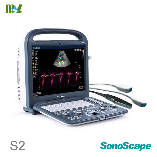
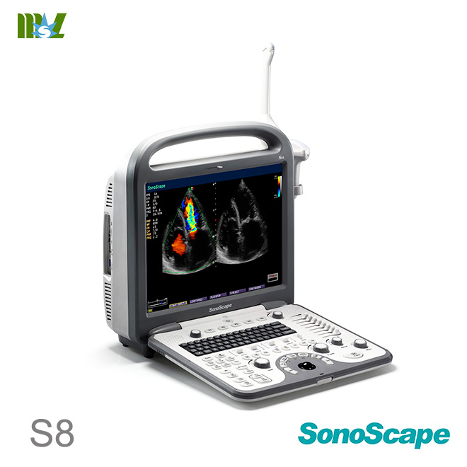
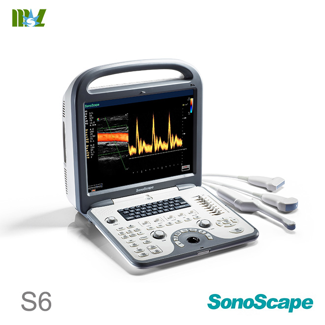
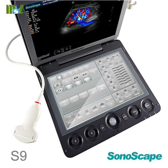
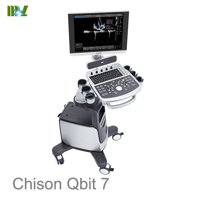
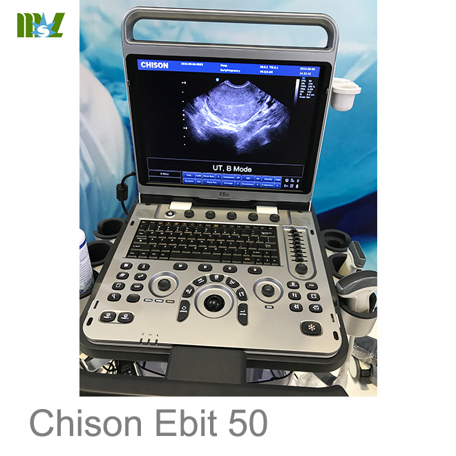
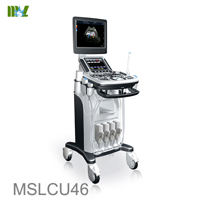
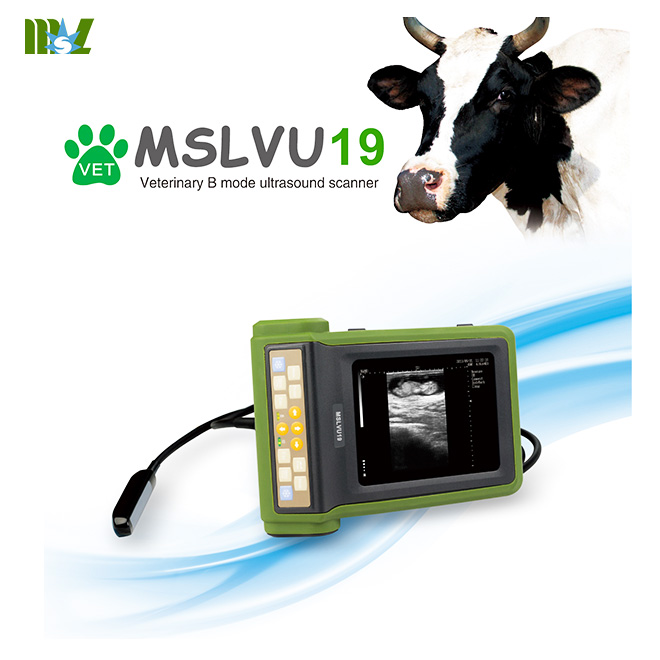
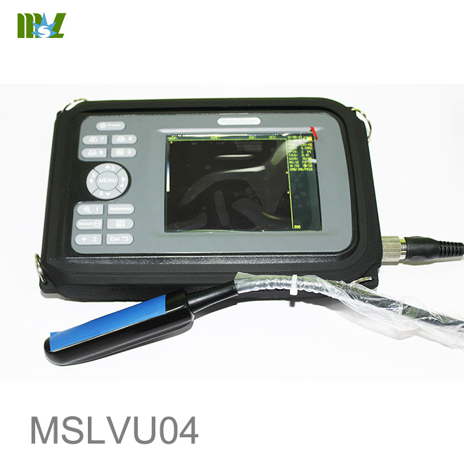
Hot sale Sonoscape ultrasound | Chison ultrasound price list


MSL Certificate
MSL Medical cooperate with DHL,FEDEX,UPS,EMS,TNT,etc.International shipping company,make your goods arrive destination safely and quickly.


 Price is 8-20% Lower Than Other