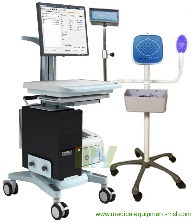
Routine EEG System | EEG instrumentation MSLEEGD
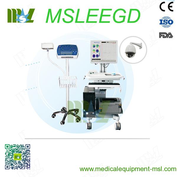
EEG instrumentation MSLEEGD Specifications
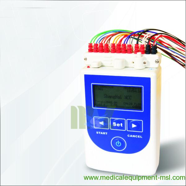
1. For MSLEEG-D Amplifier:
Input channels: 24/36/48 Monopolar EEG + 12channels Bipolar EEG,
12 channels PSG.
Power supply: Internal rechargeable lithium battery(16.8v); External charger input:
AC100~240V, output DC 19V
Connection to PC: USB (HDMI interface) cable
Operation Modes: Real-time
A/D conversion: 24 bit
Sampling Rate: 128/256/512/1024/2048/4096/8192Hz
Input impedance: ≥10MOhm
CMRR: ≥100dB
Noise: ≤0.5uVRMS
Low-pass filter: 0-120Hz
Amplitude-frequency characteristic: 1-60Hz
High-pass filter: 0.01-0.3s
Interface: USB2.0 (USB1.1), interface transfer rate: 480Mb (/ 12Mb) / s
Dimension & Weight: 300*230*60(mm); 1.65Kg (amplifier only)
2. For Stimulator (ERP Part)
2.1 A/V stimulator
2.1.1 Normal type sound stimulation parameter
Stimulation type: Short tone, Pure tone, White noise;
Stimulation mode of short tone: Left-ear, Right-ear, Binaural, Alternate;
Phase of short tone stimulation: rare wave, dense wave, alternative wave;
The maximum short tone intensity: ≥130dB
The maximum pure tone intensity: ≥120dB
The maximum white noise intensity: ≥100dB
Stimulation Frequency: 0.1~100Hz, error ≤±10%
Pure tone sound frequency: 250~7KHz, error ≤±10%
Pure tone stimulation modes: Left-ear, Right-ear, Binaural, Alternate
Target signal probability: 5%~100%, error ≤±10%
2.1.2 Video output signal
Checkerboard images, Bar grid images, vertical grid images
Grids number: 4*4~64*32
Output control: Full-screen, Half-screen (top, bottom, left, right), 1/4 screen
(I, II, III, IV quadrant display)
2.2 Current stimulator
Maximum current pulse output intensity: 100mA
Pulse output frequency: 0.1Hz ~ 100Hz
Stimulator modes: Single-stimulator mode, Dual-current stimulator mode
2.3 Photic stimulator
Stimulation frequency: 0.1~30Hz, error ≤±10%
Flash time: 1~50 minutes
Key Features & Advantages:
1. Professional EEG amplifier suitable for all standard applications of routine EEG and long term
EEG monitoring in the research and clinical areas.
2. Configurable in a number of setups according to user’s needs:
24/36/48/60 channels monopolar EEG;
24/36/48 channels monopolar EEG + 12 channels bipolar EEG;
24/36/48 channels monopolar EEG + 12 channels bipolar EEG + ERP examination with
Acoustic & Visual stimulator, consisting of three stimulation functions (sound, light and electricity)
which can be configured with special earphones and VEP monitor;
24/36/48/60 channels monopolar EEG + 12 channels polysomnography (PSG) parameters,
including: ECG (1 channel, 2 leads), Resp (1 channel), Airflow (1 channel), EOG (2 channels),
EMG (3 channels), Snore (1 channel), SpO2 (1 channel), thoracic and abdominal position (1 channel);
24/36/48/60 channels monopolar EEG + 12 channels polysomnography (PSG) parameters +
ERP examination.
3. 24 bits A/D conversion rate, with high sampling rate of up to 8 kHz.
4. High quality real-time EEG signal acquisition is transmitted via fiber optical isolation, which
shields away interference from power line and other signals, transmitting stable and reliable
measurement data at high-speed while greatly ensuring patient’s safety.
5. Amplifier can be operated either by DC power without affecting signal quality.
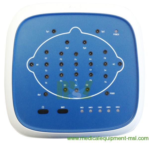
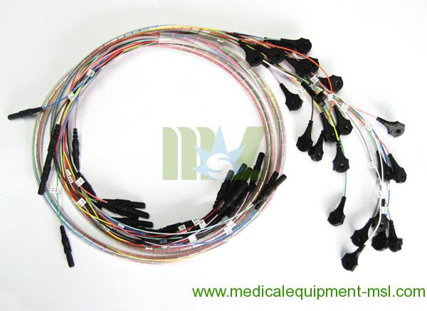
6. Head-box design to access single shielded cup/clip electrodes with touch proof
connectors (1.5mm).
7. Built-in impedance testing function and automatic calibration (for both sine wave
and square wave signals).
8. Optional video system for recording, editing and displaying video images
synchronously with EEG signals.
Software Functions:
1. Acquisition & Settings
Colors of waveforms are in accord to colors of events, and users can mark on the waveforms
with character directly; it’s available to open respiration leading sound to instruct patient's respiration
frequency in a deep respiration event;
EEG channels can be arbitrarily set up for users to customize the layout of channels during collection
process and playback analysis. This includes the choice of processing EEG waveforms with filters,
baseline and other arbitrary parameters;
Configuration chart display and data source of physical channel configuration are displayed together
at the same interface, making channel editing simpler and more intuitive;
Events markers enable user to mark timings of seizures or any abnormal wave occurrences during
recording, whereby events marked are listed and can be traced with the event localization
feature during playback;
Sound, light and electricity evoking is available during ERP data acquisition, and different
evoke methods can be applied to different patients to extract evoking waveform;
Superposition of ERP waveforms eliminates clutter and reserve EP data clearly;
Superposition of different event EP waveforms according to event's classification setting;
Same-screen, and real-time display brain tendency chart is in synchronism with EEG waveforms
acquisition. Brain tendency chart include various energy curve maps: energy curve, peak value frequency,
relative energy, absolute energy, energy peak frequency, medium frequency index,
side frequency index and coma index.
2. Replay & Analysis
EEG mapping;
EEG tendency analysis;
EEG spectral analysis;
Brain waves fast playback and fast positioning function;
Automatic spike recognition with adjustable arbitrary spike-wave parameters;
ERP data averaging function;
User can list the events marked during replay and locate each of the event markers in the
waveform. User can add, change or remove event markers based on manual observation
made during recording;
User can also review the EEG waveforms with a different combination of parameters and filters;
Various data measurement tools to measure EEG waveform, latent period and amplitude of EP;
Can display and compare up to 5 kinds of superposed data of ERP on screen;
Automatic and manual PSG analysis, RESP analysis, SpO2 analysis, EEG tendency analysis with
PSG report and RESP/PSG report;
Automatically generates EEG case reports with customizable print templates.
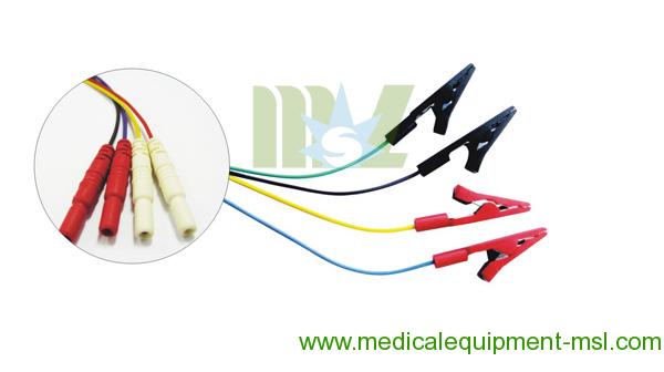
Accessories: Shielded Single disc electrode cable; Shielded Single bracket electrode cable
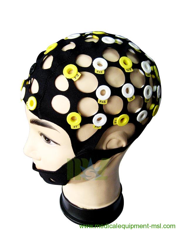
Optional accessories: Split-type EEG cap (23holes/51holes); Electrode cables for split-type EEG cap
Routine EEG System MSLEEGD Optional Parts:
1. Video System
Supported BUS Interface of video card: PCI
Power supply: AC240V 50/60Hz 2A
PAN/TILT Turn speed: 0.5°~30°/s
Night-vision survey with IR-illumination: Supported
Maximum picture resolution: 640×480
Operation System: Windows XP, Win 7
Video camera with remote control: By software.
2. Photic Stimulator:
Stimulation Frequency: 1-30Hz
General Specifications:
1. Dimension and Weight:
Type-D EEG with standard accessories: 1 carton; 510*450*230 (mm); Charge weight: 11kg
Photic Stimulator with tripod (Optional): One carton; 630*260*190 (mm); 10kg
Pentagonal stand with Photic stimulator (optional): 1 carton; 210*1000*620 (mm); Charge weight: 25kg
Video system (optional): 1 carton; 450*300*300 (mm); Charge weight: 7kg
2. Environment:
Temperature: ±10°C ~+50°C
Relative humidity: 30%~80%
Power supply: AC100—240V (Host computer system)
Amplifier: DC19V±
3. Quality System:
ISO13485: 2003; CE (93/42/EEG)


 Price is 8-20% Lower Than Other






![{pr0int $v['title']/}](https://medicalequipment-msl.com/upload/img/20141106/201411061510127441.jpg.jpg)
![{pr0int $v['title']/}](https://medicalequipment-msl.com/upload/img/20150331/201503311430334326.jpg.jpg)
![{pr0int $v['title']/}](https://medicalequipment-msl.com/upload/img/20161130/201611301725306855.jpg.jpg)
![{pr0int $v['title']/}](https://medicalequipment-msl.com/upload/img/20150611/201506111820203873.jpg.jpg)
![{pr0int $v['title']/}](https://medicalequipment-msl.com/upload/img/20130722/201307221809154861.jpg.jpg)
![{pr0int $v['title']/}](https://medicalequipment-msl.com/upload/img/20130718/201307181746235998.jpg.jpg)
![{pr0int $v['title']/}](https://medicalequipment-msl.com/upload/img/20161201/201612011821596048.jpg.jpg)
![{pr0int $v['title']/}](https://medicalequipment-msl.com/upload/img/20150610/201506101156246760.jpg.jpg)
![{pr0int $v['title']/}](https://medicalequipment-msl.com/upload/img/20190614/201906141354248163.jpg.jpg)
![{pr0int $v['title']/}](https://medicalequipment-msl.com/upload/img/20150626/201506261614056119.jpg.jpg)
![{pr0int $v['title']/}](https://medicalequipment-msl.com/upload/img/20130718/201307181632356998.jpg.jpg)
![{pr0int $v['title']/}](https://medicalequipment-msl.com/upload/img/20130722/20130722173929721.jpg.jpg)
![{pr0int $v['title']/}](https://medicalequipment-msl.com/upload/img/20150626/201506261539406148.jpg.jpg)
![{pr0int $v['title']/}](https://medicalequipment-msl.com/upload/img/20161202/201612021531037589.jpg.jpg)
![{pr0int $v['title']/}](https://medicalequipment-msl.com/upload/img/20150611/201506111604066010.jpg.jpg)
![{pr0int $v['title']/}](https://medicalequipment-msl.com/upload/img/20150619/201506191439309321.jpg.jpg)
![{pr0int $v['title']/}](https://medicalequipment-msl.com/upload/img/20161201/201612011835218235.jpg.jpg)
![{pr0int $v['title']/}](https://medicalequipment-msl.com/upload/img/20130718/20130718174229220.jpg.jpg)
![{pr0int $v['title']/}](https://medicalequipment-msl.com/upload/img/20150626/201506261722125019.jpg.jpg)
![{pr0int $v['title']/}](https://medicalequipment-msl.com/upload/img/20150611/201506111035316960.jpg.jpg)
![{pr0int $v['title']/}](https://medicalequipment-msl.com/upload/img/20130817/201308171711045217.jpg.jpg)
![{pr0int $v['title']/}](https://medicalequipment-msl.com/upload/img/20131021/201310211505084105.jpg.jpg)
![{pr0int $v['title']/}](https://medicalequipment-msl.com/upload/img/20150626/201506261429219782.jpg.jpg)
![{pr0int $v['title']/}](https://medicalequipment-msl.com/upload/img/20130718/201307181750089757.jpg.jpg)
![{pr0int $v['title']/}](https://medicalequipment-msl.com/upload/img/20130722/201307221806554299.jpg.jpg)


