


 Price is 8-20% Lower Than Other


bovine ultrasound machines plays an important role in the examination of the lumbar spine pain.Especially in the lumbar artery exploration.
Maintain adequate blood supply due to the normal function of the lumbar spine is very important:for capillary blood from the bone between the lumbar artery;disc of the student movement from the bone between the capillary network;destroy these blood vessels and blood flow can lead to disc blood for other important structural barriers to yield the corresponding disease state.Some scholars believe that the abdominal aorta of atherosclerotic plaques and other diseases can cause arterial transportation waist reduced,thereby inducing disc degeneration and low back pain,the study of vascular hemodynamic waist help clinicians better understand them and disc degeneration,low back pain and other related properties.
Currently waist vascular hemodynamic data on the normal population,and the population is still not reported back pain,and color Doppler ultrasound as a non-invasive examination methods assisted hemodynamic monitoring,which is currently in clinical applications have been increasingly the more extensive,and its hemodynamic measurements have confirmed the reliability of data for clinical research institute.Recently,scholars from Oxford University orthopedic applications on color Doppler ultrasound in the detection of blood flow dynamics in patients with low back pain,a technical report,and compare the normal population and in patients with low back pain associated hemodynamic data and conclusions published in Spine magazine.
Study is divided into two groups,one group with low back pain,low back pain for three months,in the past by MRI,patients diagnosed with lumbar disc degeneration presence of 64 patients ( 35 females and 29 males,age range 21 - 82 years;mean age of 50 ± 16.5 years);normal group,back pain does not occur within the previous June had not received medical treatment because of back pain,a total of 30 cases (13 females and 17 males,age range 19 - 82 years,mean age 41 ± 18.5 years).
Radiographic evaluation methods: carry blood from the rear of the lumbar spine to detect.Start L5-S1 begin to determine the fifth lumbar artery and sacral artery,then up probe to determine 4th - 1st lumbar artery ( Figure 1 ).
Figure 1:Color Doppler ultrasound probe(color ultrasound machine),more visible vessel branches emanating from the extension of the lumbar facet joints spinous
Get bilateral lumbar artery hemodynamic data,the use of Doppler ultrasound-assisted software records the peak systolic velocity (peak systolic velocity,PSV,Figure 2 ).A total of 12 data,each side 6.Changes produced in order to avoid the situation of human physiological impact on the statistical data,the PSV values standardized calculation PSVR value (PSV ratio)=lumbar artery PSV values aortic PSV values.
Figure 2:Color Doppler ultrasound showed the traffic branch.
The results showed that :
The normal population,lumbar artery PSV values,showing a gradual increasing trend from the first to the fourth lumbar arteries lumbar artery,while the smallest in the fifth lumbar artery PSV values;PSV values sacral artery has increased over the fifth lumbar artery,but Less than a fourth lumbar artery;there were significant differences (p <0.001) between the segmental arteries compare PSV,but about bilateral comparison was no significant difference (p=0.09),as shown in Table 1.
Table 1: lumbar artery PSV
Aortic PSV shown in Table 2,the probe found no evidence of aortic atherosclerosis;although PSV's statistical difference (p <0.01) exists between the low back pain group and the normal group,but these differences are not of clinical significance because the two groups of patients with aortic PSV were within the normal range.
Table 2:PSV value aorta
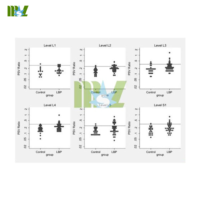
Low back pain group and normal group PSVR comparison: Table 3 shows a comparison of the data back pain group and the normal group PSVR data area.Two aspects of the same trend increase in PSVR:L1-L4 increases,L5 reduced to a minimum value,S1 rise again,there is a significant difference (p <0.001) between segments comparison;between back pain group segments PSVR value than the normal population set high (p <0.001),between groups and segments no significant correlation values (p=0.9).Table 4 shows the PSV 95% confidence interval,Figure 3 shows the various groups PSVR values on different segments.
Table 3: PSVR value distribution
Table 4: PSV 95% confidence interval
Figure 3:Each group PSVR values on different segments of
Conclusions:PSVR value of low back pain patients was significantly higher than the normal population,the characteristics and Doppler ultrasound vascular congestion(cheap ultrasound machine) when the change is similar to changing conditions and dynamics of the relationship between the pathogenesis of low back pain in patients with low back pain blood still needs further clarification.
- Home
- Ultrasound
- Sonoscape ultrasound
- 4D ultrasound machine
- 3d Ultrasound Machine
- Wireless ultrasound
- Portable Ultrasound Machine
- Color Ultrasound Machine
- Bone Densitometer Machine
- Handheld Ultrasound
- Veterinary Ultrasound
- Trolly ultrasound machine
- Digital Ultrasound Machine
- Chison ultrasound
- Home Ultrasound Machine
- Mindray ultrasound
- Fetal Doppler
- Medical Printer
- Laboratory
- Automated blood analyzer
- Dry Chemistry Analyzers
- Biochemistry analyzer
- Veterinary blood analyzer
- Immunoassay analyzer
- Blood gas analyzer machine
- Urinalysis machine
- Rayto IVD
- Microplate readers
- Mindray analyzer
- Testing equipment
- Blood cell analyzer
- Portable blood analyzer
- Blood coagulation analyzer
- Blood pressure analyzer
- Handheld blood analyzer
- Radiology
- Surgery
- Veterinary
- Beauty
- ENT treatment
- Ophthalmology
- Contact


