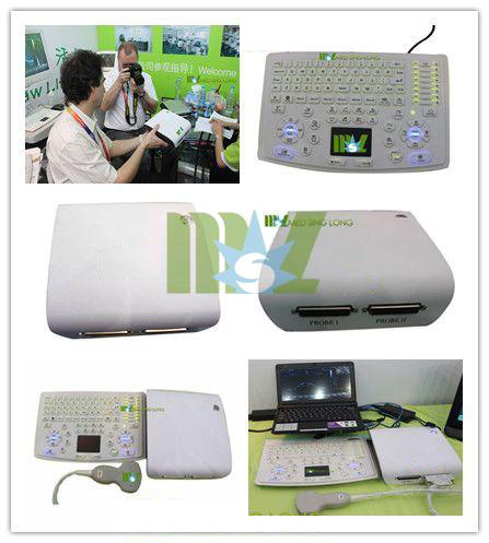
The portable veterinary ultrasound scanner solved the problem of observation about corpus luteum during different estrous stages.Formerly people get ovary from the slaughterhouse and then start direct and surface observation.They determine the corpus luteum's stages by the outer color (red,yellow and white),or cut the ovary to verify it by the internal situation.Guangzhou Medsinglong Global Group LTD introduces how to determine the maturity of the cow's ovum with a veterinary ultrasound scanner to inspect the corpus luteum.
Firstly,use a veterinary ultrasound scanner to observe the follicle and the uterus mucus when oestrus.Under the ultrasound imaging,the follicular fluid is hypoechoic fluid dark area and the hypoecho in the uterine horn is the estrus mucus
Secondly,use the veterinary ultrasound scanner to observe the new corpus luteum after ovulation.
Mostly,the new corpus luteum is with a fluid chamber and the chamber is large.In the ultrasound imaging,it's medium or low density organization.

Thirdly,use the veterinary ultrasound scanner to observe new corpus luteum and follicle.
Evaluation of follicles under the state of this new corpus luteum: follicle of 1.79CM used to be a cyst and is of aging or stationary state currently.
Fourthly,use the veterinary ultrasound scanner(Portable Ultrasound scanner box) to observe the corpus luteum the third day after ovulation.The liquid chamber within the corpus luteum is large and there is are rules of close loop medium echo surrounds the liquid cavity
Fifthly,use the veterinary ultrasound scanner to observe the corpus luteum the sixth day after ovulation.
The liquid chamber shrinks and the circular echo becomes more obvious.It's important to note that the liquid chamber of early corpus luteum isn't all round or neatly edged.While it trends to develop into round chamber.
Sixthly,use the veterinary ultrasound scanner to observe the corpus luteum the seventh day after ovulation.
The liquid is circular and the annular zone is regular and clear,at the same time the small follicles develop.
Seventhly,use the veterinary ultrasound scanner(Ultrasound box) to observe the corpus luteum the eightth day after ovulation.
The liquid Chamber further shrinks and the annular zone further enlarges.The small follicles develop.The diameter of the corpus luteum is 3.46CM,it's much larger than the graafian follicle's diameter 8 days ago.There is no unified dimension to measure the liquid chamber 6 days,7 days and 8 days after ovulation.
The state of corpus luteum in the cow's ovary is always ignored by people.To accurately determine the corpus luteum growth stage or days is of great help to the general evaluation of ovary and the understanding of the follicle.General evaluation of ovary combines with track the follicle ovulation time,can help us to decide whether to transplant and the time to transplant.
- Home
- Ultrasound
- Sonoscape ultrasound
- 4D ultrasound machine
- 3d Ultrasound Machine
- Wireless ultrasound
- Portable Ultrasound Machine
- Color Ultrasound Machine
- Bone Densitometer Machine
- Handheld Ultrasound
- Veterinary Ultrasound
- Trolly ultrasound machine
- Digital Ultrasound Machine
- Chison ultrasound
- Home Ultrasound Machine
- Mindray ultrasound
- Fetal Doppler
- Medical Printer
- Laboratory
- Automated blood analyzer
- Dry Chemistry Analyzers
- Biochemistry analyzer
- Veterinary blood analyzer
- Immunoassay analyzer
- Blood gas analyzer machine
- Urinalysis machine
- Rayto IVD
- Microplate readers
- Mindray analyzer
- Testing equipment
- Blood cell analyzer
- Portable blood analyzer
- Blood coagulation analyzer
- Blood pressure analyzer
- Handheld blood analyzer
- Radiology
- Surgery
- Veterinary
- Beauty
- ENT treatment
- Ophthalmology
- Contact


 Price is 8-20% Lower Than Other


