Newest touch screen laptop ultrasound machine MSLCU29 for sale
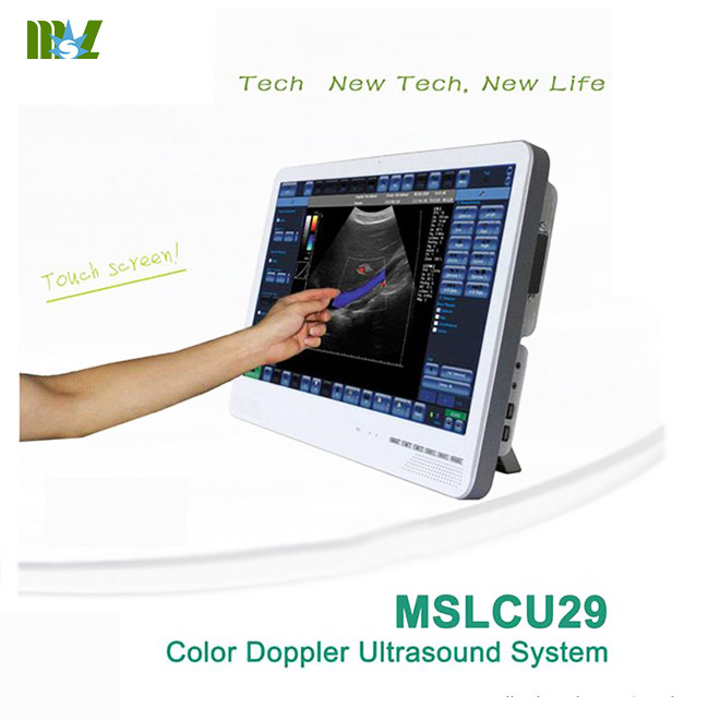
touch screen laptop ultrasound machine MSLCU29 for sale Feature:
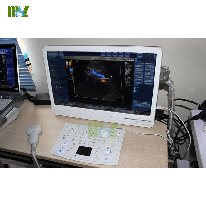
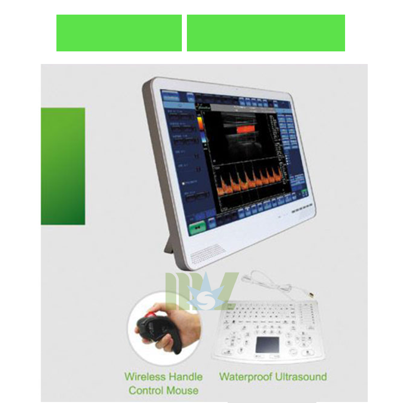
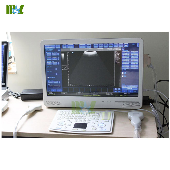
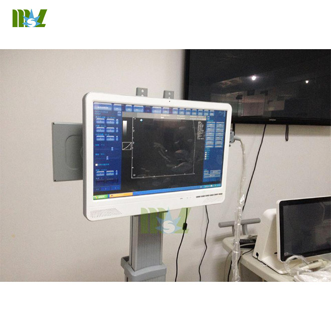
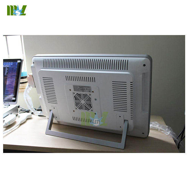
-22 inch color LED Touch Screen
-With multiple probes to fit different uses and budgets
-Imaging Mode: B, BB, M, CD, PWD, CWD, DirPwr, Pwr
-High Definition Multiple Color Doppler mode imaging, include supports most Doppler imaging and advanced imaging
processing for cardiac, vascular access, and OB diagnostics.
-Can be equipped in addition with 3D and panoramic image processing for volume reconstruction, visualization,
segmentation, and measurement.
-Can install and run all applications with Microsoft? Windows? including patient management systems for better follow up,
as well as network tools to access PACS data
-Large volume store and preview, edit records (image, voice comments, cineloop video) by hardisk, USB flash disk, DVD
-Triplex Display: Real-time triplex display B/ Color Doppler/ Pulsed Spectral Doppler (Three TGCs can be adjusted respectively)
-THI (Tissue Harmonic Imaging) technology: Suppress speckle noise, brighten image and improve the image quality
-One-key Optimization: One key for eight-parameter adjustment, easy image optimization.
-Powerful Measurement Software: Versatile clinic-oriented measuring software package
-Image Management: Versatile image format/ large capacity cineloop storage/ preview/ edit
-Can maintain and update by internet
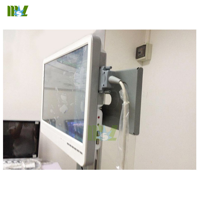
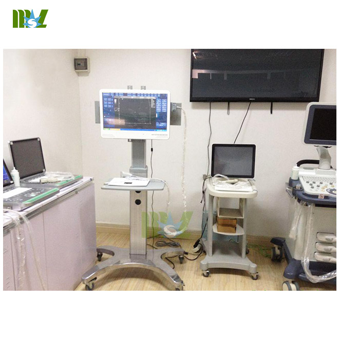
-Space saving by the integration of color doppler ultrasound scanner and office computer, and can either lay
on a desk or be wall mounted.
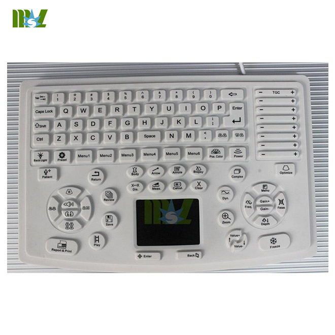
-Can be controlled either through a regular PC keyboard and a mouse, or a dedicated ultrasound keyboard.
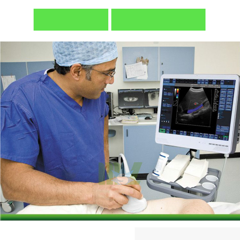
-Except general use, special suit for use in operation room or ambulances
-2 probe sockets
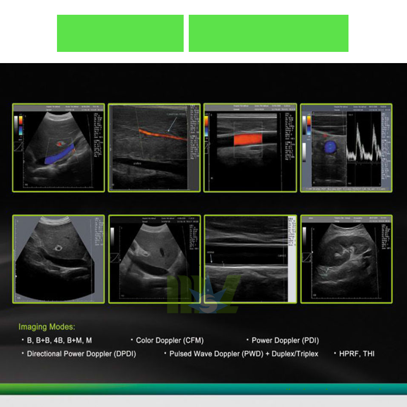
laptop ultrasound machine MSLCU29 for sale Imaging Modes
•B
•B+B
•4B
•B+M
•M
•B-steer
•Compound + Trapezoid
•Color Doppler (CFM)
•Power Doppler (PDI)
•Directional Power Doppler (DPDI)
•Pulsed Wave Doppler (PWD)
•B+PWD (Duplex)
•B+CFM/PDI/DPDI+PWD (Triplex)
•High Pulse Repetition Frequency (HPRF)
•Tissue Harmonic Imaging (THI)
Ultrasound imaging
•ultrasound image size: automatically adjustable to screen resolution
•gray scale: 256
•color scale: 256
•full motion and full size real-time ultrasound imaging, up to 120 fps (depends on selected scan depth, scan angle, focus mode,
High Line Density setting, computer speed)
•cineloop recording/play: several thousands frames (depends on computer memory size and scan mode)
•zoom mode: from 60% to 600% in all modes (Scan, Freeze, B, B+B, 4B, Doppler modes, M-zoom, cineloop and etc)
•viewing area variable for frame rate maximizing: 6 steps
•thumbnail mode: up to 32 images
•"Freeze" mode
•"Auto Freeze" mode
Scanning Method
•electronic linear
•electronic convex
•electronic microconvex
•scanning depth: 2-30 cm
Transducers
•convex, micro-convex, linear, transvaginal
•from 2,0 MHz to 12,0 MHz
•multifrequency
•automatic transducer recognition
Color Doppler
•PRF variable: 0.5-10 kHz
•wall filter settings: 3 steps (5%, %10%, 15% PRF)
•gain control: 50 dB
•angle steering for linear transducers: ±10°
•real-time spatial filter: 4 values
•CFM palette: 10 maps
•PDI palette: 11 maps
•B/Color priority control
•color threshold control
•CFM baseline control
•Doppler frequency selection: 2 frequencies / each transducer
•color frame averaging: 8 values
•Transparent Color Mapping (TCM): 10 values
Automatic Image Optimization
•single click auto adjustment:
•B-image: gain, dynamic range, TGC sliders
•Color Doppler: CFM/PDI/DPDI gain
•Pulsed Waved Doppler: baseline, invert, PRF
Pulsed Wave Doppler
•PRF variable: 1-15 kHz
•wall filter settings: 16 steps (2.5%-20% PRF)
•gain control: 50 dB
•angle steering for linear transducers: ±10°
•real-time trace line with automatic calculation of spectrum parameters
•stereo sound: volume control
•PWD palette: 12 maps
•Doppler frequency selection: 2 frequencies / each transducer
ultrasound machine MSLCU29 for sale Focusing
digital transmit focusing
multi focus mode:
transmit/receive focusing
programmable focus area presets
dynamic focus mode:
transmit variable focus
dynamic receive focus
Newest touch screen laptop ultrasound machine MSLCU29 Processing
High Line Density scan mode for better resolution
TGC Control, 5-10 sliders (customizable) 40 dB
dynamic range: 120 dB, 8 values
overall gain control
M - mode sweep speed control
acoustic power control
variable frame averaging
brightness, contrast
advanced gamma control: 8 fixed curves, 8 user defined (custom)
scan direction, rotation, up-down controls
negative / positive control
bi-linear interpolation
echo enhancement control
noise rejection function
speckle reduction and structure improvement PureView: 8 algorithms
touch screen laptop ultrasound machine MSLCU29 Functions
mouse / trackball / keyboard operation
anatomical icons with transducer position indicator
direct e-mail sending with image or video attachment via Internet
DICOM file push to server
printing on system printer
unlimited programmable presets for clinically specific imaging
TV output via computer display adapter
File Formats
image and video save / load
AVI
JPG
BMP
PNG
TIF
DCM (DICOM uncompressed)
DCM (DICOM-JPEG RGB/YBR)
DCM (DICOM-JPEG RGB/YBR Video)
TPD (Picture Data)
TVD (Video Data)
DICOM
Verification SCU
Modality Worklist (MWL) SCU
Modality Performed Procedure Step (MPPS) SCU
Store SCU (images, cines)
Print SCU (grayscale, color)
laptop ultrasound machine MSLCU29 Interface Customization
the set of predefined skin schemes for software interface
the set of predefined buttons images
Multilanguage support, languages:
Chinese
English
German
Italian
Korean
Lithuanian
Magyar
Polish
Romanian
Russian
Spanish
B+M, B+PW layout position, size
ultrasound area size
font size
ultrasound machine MSLCU29 General Measurements and Calculations
B and Color Doppler mode general measurements and calculations
Distance
Length (method: 1 trace)
Area, Circumference (methods: 1 ellipse, 1 trace, 1 distance)
Volume (methods: 1 distance, 2 distances, 3 distances, 1 ellipse)
Angle (methods: 2 distances, 3 distances)
Stenosis % (methods: 2 distances, 2 ellipse or trace areas)
A/B Ratio (methods: 2 distances, 2 ellipse or trace areas, 2 ellipse or trace circumferences)
M mode general measurements and calculations
Distance, Time, Velocity
Heart Rate (methods: 1 beat, 2 beats)
Stenosis % (method: 2 distances)
A/B Ratio (methods: 2 distances, 2 times, 2 velocities)
PW mode general measurements and calculations
One-point PW measurements and calculations:
Velocity
Pressure Gradient (PG)
Two-points PW measurements and calculations:
Velocities difference
Pressure Gradients (PG) difference
Time interval
Acceleration
Resistivity Index (RI)
Heart Rate (methods: 1 beat, 2 beats)
Velocity minimum and maximum
Pressure Gradient (PG) minimum and maximum
Trace-based PW measurements and calculations:
Trace Time
Trace Velocity min, max, mean
Trace Pressure Gradient (PG) min, max, mean
Velocity Time Integral (VTI)
Pulsatility Index (PI)
A/B Ratios of one-point PW measurements:
Velocities A/B Ratio
Pressure Gradients (PG) A/B Ratio
A/B Ratios of two-point PW measurements:
Velocity differences A/B Ratio
Pressure Gradient (PG) differences A/B Ratio
Time differences A/B Ratio
Accelerations A/B Ratio
Resistivity Indexes A/B Ratio
A/B Ratios of trace-based PW measurements:
Velocity means A/B Ratio
Pressure Gradient (PG) means A/B Ratio
Pulsatility Indexes A/B Ratio
Velocity Time Integrals A/B Ratio
MSL Newest touch screen laptop ultrasound machine MSLCU29 PW mode Human Cardiology measurements and calculations
Left Ventricle: LVOT Diam, LVOT VTI, LVOT Vmax, SV (Stroke Volume), SI (Stroke Volume Index), CO (Cardiac Output),
CI (Cardiac Index), dP:dt (Delta Pressure : Delta Time), MPI (Left Ventricle Myocardial Performance Index)
Mitral Valve: MVA(PHT) (Mitral Valve Area using Pressure Half Time), MVA using Continuity Equation (LVOT Diam, MV VTI;
LVOT Diam, MV Vmax), dP:dt, E/A ratio
Aortic Valve: AVA (Aortic Valve Area) using Continuity Equation (LVOT Diam, AV VTI; LVOT Diam, AV Vmax),
AVI (Aortic Valve Index), DPI (Dimensionless Performance Index), AV PHT (Aortic Valve Pressure Half Time)
Right Ventricle: RVOT Diam, RVOT VTI, RVOT Vmax, dP:dt, RV MPI (Right Ventricle Myocardial Performance Index),
MPAP (Mean Pulmonary Artery Pressure)
Tricuspid Valve: TVA (Tricuspid Valve Area) using Continuity Equation (RVOT Diam, TV VTI; RVOT Diam, TV Vmax);
TV E/A ratio, TV PHT
Pulmonic Valve: PVA (Pulmonic Valve Area) using Continuity Equation (RVOT Diam, PV VTI; RVOT Diam, PV Vmax),
PVI (Pulmonic Valve Index), DPI (Dimensionless Performance Index), PV PHT (Pulmonic Valve Pressure Half Time)
Pulmonary Vein; Hepatic Vein
Shunts: Qp:Qs (Pulmonary-Systemic Flow Ratio)
MSLtouch screen laptop ultrasound machine MSLCU29 Human Measurements and Calculations Packages
Human General calculations package
Measurements: the same as general measurements of different modes (B, M, PW)
Calculations: BSA via Height and Weight, BSA via Weight, Hip Angles (α, β), Femoral Head Coverage (FHC)
PW mode calculations: HR, SV using Flow Area, SV using Flow Diameter, SI, CO, CI, Area calculation using Continuity Equation (methods: Area and VTI, Area and Velocity, Diameter and VTI, Diameter and Velocity, Velocity Ratio (S/D, D/S), dP:dt, Flow Volume (methods: Diameter, Area), PHT, MVA
Human Obstetrics / Gynecology (OB / GYN) calculations package
Measurements: LMP (entered or from calendar), AC, BPD, FL, HC, FTA, AAPD, ATD, TAPD, TTD, CRL, GS, HL, TL, UL,
OFD, BOD, Cereb, Clav, Rad, AFI, FHR
Estimated date of birth (EDD) calculations: EDD(LMP), EDD(GA), EDD(AUA)
Ratios: FL / AC, FL / HC, FL / BPD, HC / AC, CI
Estimated Fetal Weight (EFW) calculations: EFW(AC), EFW(AC,BPD), EFW(AC,FL), EFW(AC,HC), EFW(AC,HC,BPD),
EFW(FL), EFW(AC,FL,HC), EFW(AC,BPD,FL), EFW(AC,BPD,FL,HC), EFW(BPD,FL,FTA), EFW(BPD,ATD), EFW(BPD,TTD),
EFW(BPD,TAPD,TTD), EFW(BPD,FL,TAPD,TTD)
Selected EFW values are used for calculation of Average EFW
Gestational Age (GA) calculations: GA(AC), GA(BPD), GA(CRL), GA(FL), GA(GS), GA(HC), GA(HL), GA(OFD), GA(TL),
GA(UL), GA(HC/AC), GA(FTA),GA(ATD), GA(TAPD), GA(TTD), GA(BOD), GA(Cereb), GA(Clav)
Fetal Growth estimation (trending): AC(GA), BPD(GA), CRL(GA), FL(GA), GS(GA), HC(GA), HL(GA), OFD(GA), TL(GA),
UL(GA), [FL/AC](GA), [FL/HC](GA), [HC/AC](GA), EFW(GA), AAPD(GA), ATD(GA), TAPD(GA), TTD(GA), BOD(GA), Cereb(GA),
Rad(GA), Clav(GA), AFI(GA), FHR(GA)
Selected Growth Tables are visualized as Fetal Growth Curves
Software supports unlimited number of user-defined Growth Tables
Human Gynecology (GYN)
Measurements: length, height, width of uterus, cervix, ovaries, renals, follicles
Calculations: volumes of uterus, cervix, ovaries, renals, follicles
Human Abdominal exam measurements and calculations
Liver: Volume (CC, AP, LL diameters)
Gallbladder: Volume, Wall Thickness, Extrahepatic Bile Duct (EBD), Common Bile Duct (CBD), Common Hepatic Duct (CHD)
Pancreas: Head Diameter, Body Diameter, Tail Diameter, Pancreatic Duct Head, Pancreatic Duct Body
Spleen: Volume (length, width, thickness)
Gastrointestinal Tract: Appendix Wall Thickness, Appendix Diameter, Bowel Wall Thickness (at Stomach, Small Bowel, Large Bowel)
Urinary Bladder: Volume (length, height, width)
Right / Left Kidney: Volume (length, height, width), Pelvis Diameter
Human Urology
Measurements: length, height, width of kidneys, bladder, prostate, testes
Calculations: volumes of kidneys, bladder, prostate, testes; RUV (Residual Urine Volume)
Human Endocrinology
Measurements: length, width, thickness of thyroid lobes
Calculations: volumes thyroid lobes; volume of thyroid
Human Vascular exam measurements and calculations
Distance and area -based stenosis calculations at left (right) Subclavian, CCA (Common Carotid Artery), Bulb,
ICA (Internal Carotid Artery),
ECA (External Carotid Artery), Vertebral vessels at proximal, middle, distal locations
PSV/EDV (Peak Systole Velocity / End Diastole Velocity) ratios for each vessel and location
Ratios of velocities ICA PSV/CCA PSV, ICA EDV/CCA EDV, ICA PSV/CCA EDV, ECA PSV/CCA PSV, ECA EDV/CCA EDV,
ECA PSV/CCA EDV at Rt. (Lt.) Prox. (Mid., Dist.) locations
Human Cardiology
Software supports the following measurements of Left Ventricle, Aortic Valve,
Left Atrial: IVSd (Interventricular Septal Thickness, diastole), LVIDd (Left Ventricle Internal Diameter, diastole),
LVPWd (Left Ventricle Posterior Wall Thickness, diastole), AOd (Aortic Root Dimension, diastole),
IVSs (Interventricular Septal Thickness, systole), LVIDs (Left Ventricle Internal Diameter, systole), LVPWs
(Left Ventricle Posterior Wall Thickness, systole), LADs (Left Atrial Dimension, systole).
Calculations: HR (Heart Rate), BSA (Body surface Area), Left ventricle volume (methods: Cubed, Teichholz, Gibson,
Simpson’s LVAM-LVAP, Simpson’s single plane, Simpson’s biplane, Bullet, Ellipsoid single plane, Ellipsoid biplane),
SV (Stroke Volume), SI (Stroke Volume Index), EF (Ejection Fraction), CO (Cardiac Output), CI (Cardiac Index),
STIVS (Interventricular Shortening), FS (Fractional Shortening), STPW (Posterior Wall Shortening),
LVM (Left Ventricle Cardiac Mass), CMI (Cardiac Mass Index), LA/AO Ratio
Human/Veterinary OB Gyn packages: software supports unlimited number of user-defined GA tables,
selected GA values are used for calculation of Average GA (Average Ultrasound Age - AUA).
Human/Veterinary Cardiology measurements package automatically displays hint images
that show where and how appropriate measurements must be performed.
MSL laptop ultrasound machine MSLCU29 Veterinary Calculations Packages
Canine OB
Measurements: GS, CRL, HD, BD
Gestational Age (GA) calculations: GA(BD), GA(CRL), GA(GS), GA(HD)
Feline OB
Measurements: HD, BD
Gestational Age (GA) calculations: GA(BD), GA(HD)
Ovine OB
Measurements: CRL
Gestational Age (GA) calculations: GA(CRL)
Bovine OB
Measurements: BD, CRL, HD, UD
Gestational Age (GA) calculations: GA(BD), GA(CRL), GA(HD), GA(UD)
MSL ultrasound machine MSLCU29 Veterinary Calculations Packages
Equine OB
Measurements: AOD, BPD, CRL, EOD, GS
Gestational Age (GA) calculations: GA(AOD), GA(BPD), GA(CRL), GA(EOD), GA(GS)
Llama OB
Measurements: BPD
Gestational Age (GA) calculations: GA(BPD)
Goat OB
Measurements: BPD
Gestational Age (GA) calculations: GA(BPD) for different species
Animal Cardiology
Left Ventricle, Aortic Valve, Left Atrial measurements: IVSd, LVIDd, LVPWd, AOd, IVSs, LVIDs, LVPWs, LADs, ET
Calculations: HR, LV volume (Cubed, Teichholz, Gibson), SV, EF, CO, STIVS, FS, STPW, LA/AO, VCF
Standard Configuration:
-Host 1 unit
-Convex probe 1 pcs
-Lienar or Trans-vaginal probe 1 pcs
-Computer keyboard 1 pcs
MSL laptop ultrasound machine MSLCU29 for sale Optional:
Linear, Trans-vaginal, Micro-convex, Trolley, Printer
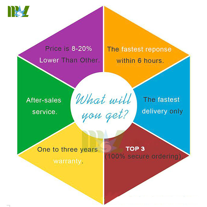
what will you get?
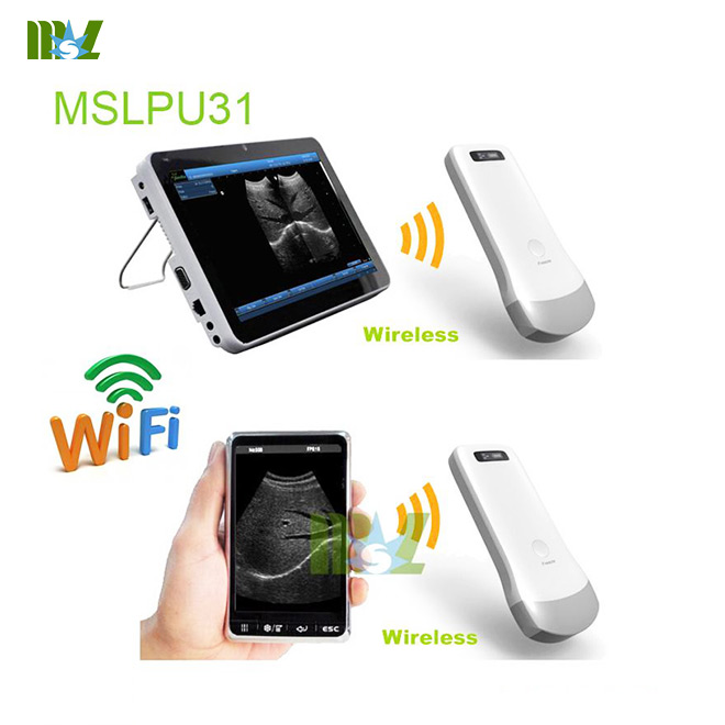
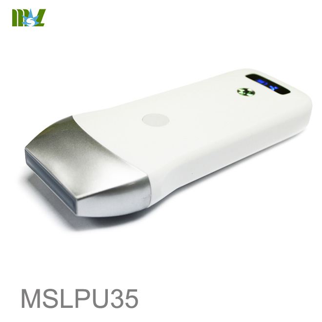
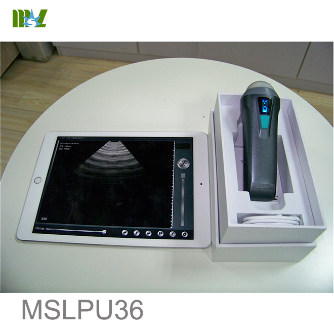
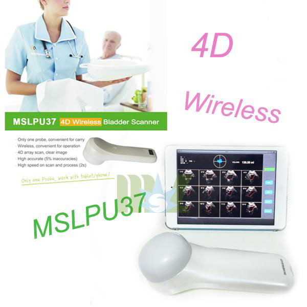
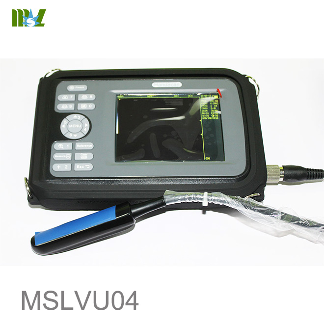
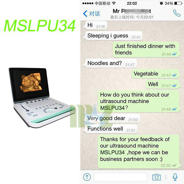
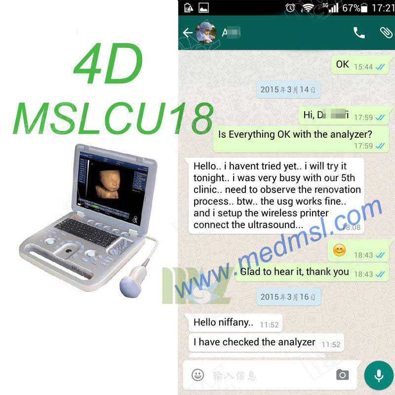
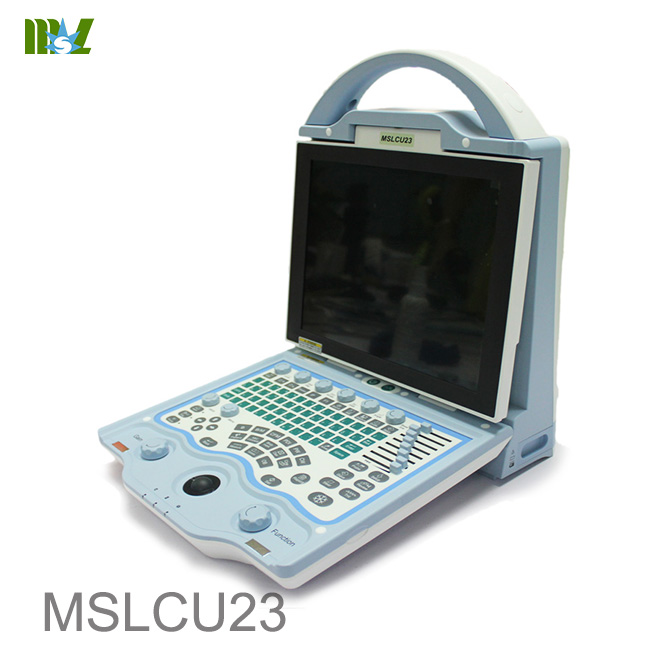
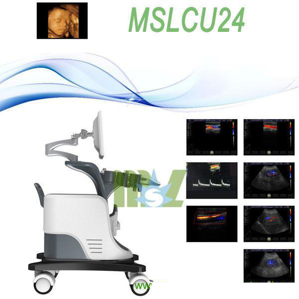
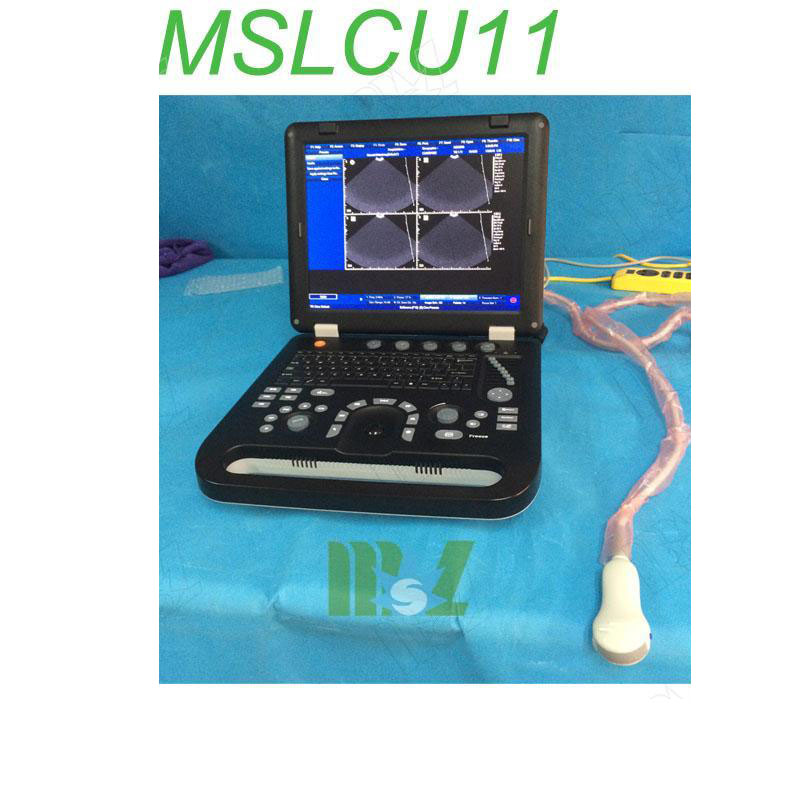
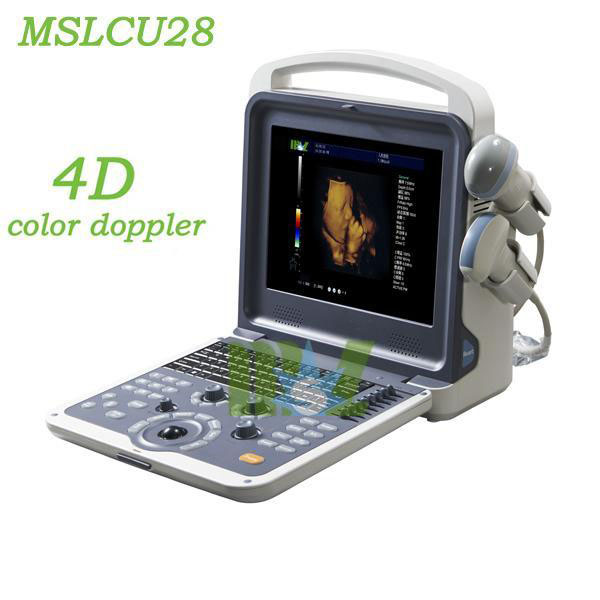
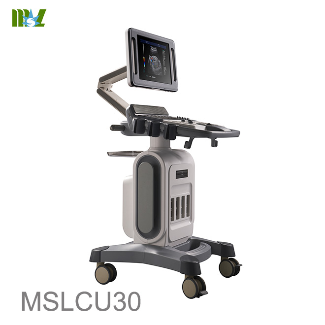
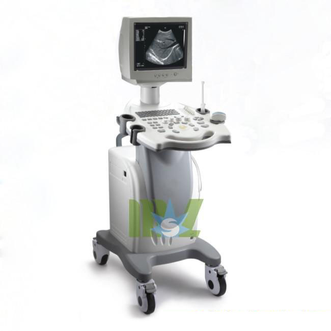
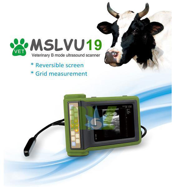
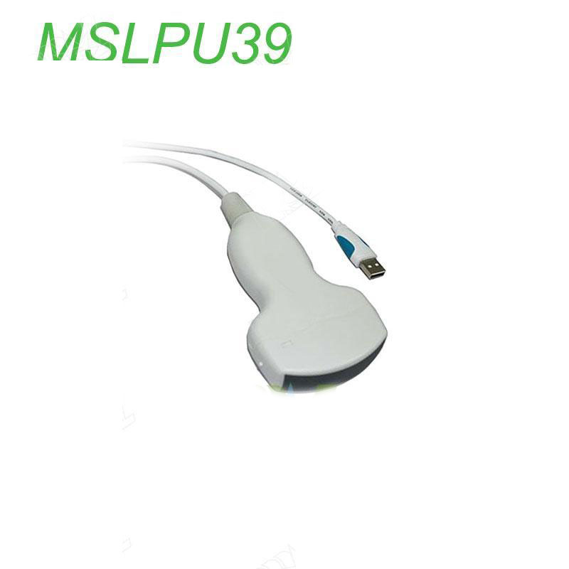
Other Hot sale ultrasound machine recommend
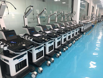
MSL TEAM picture


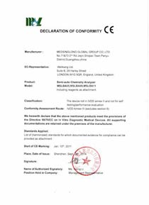
MSL Certificate
MSL Medical cooperate with DHL,FEDEX,UPS,EMS,TNT,etc.International shipping company,make your goods arrive destination safely and quickly.


 Price is 8-20% Lower Than Other