B-Mode
B-mode ultrasound is one of the most commonly used operation modes of a clinical ultrasound machine. As explained earlier, ultrasound is a reflection or scattering based imaging modality, and the sophisticated generation of a sound wave allows the focusing of the sound to a specific location. Each transmission yields one scan line around the targeted focal point. If only one focal point is selected, one scan line extends over the total depth range, which is user defined in the current imaging settings. In order to record a full image frame using a linear array, the imaging software of the scanner electronically moves the active aperture of the array across the physical aperture to transmit and receive at a given line density.
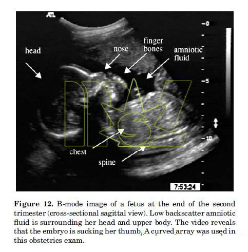
Typically, hundreds of scan lines are generated this way and displayed on a monitor. Figure 12 shows the cross-sectional sagittal (front to back, vertical slice) image of a fetus in utero. She is sucking on her thumb, as real-time video reveals. On the left of Fig. 12 one can see the head and the strong reflection of the skull bone. The very left side of the image is black, an artifact that could be due to maternal bowel gases that scatter the sound away from the transducer. The remainder of the skull is clearly visible from the forehead to the chin and from the back of the head to the neck area. Bones reflect sound waves well and result in a bright signal in the image. The black surroundings of the fetus are regions of amniotic fluid, which does not scatter sound due to the homogeneous nature of the fluid.

In front of the mouth, one can see the hand of the fetus. Once again the bones of the hand, namely, the knuckles, are pronounced since they scatter more ultrasound than the soft tissue of the hand. In the same fashion one can see the reflections of the spine.


 Price is 8-20% Lower Than Other


