Factory direct sales ecografo Chison Q9 price list
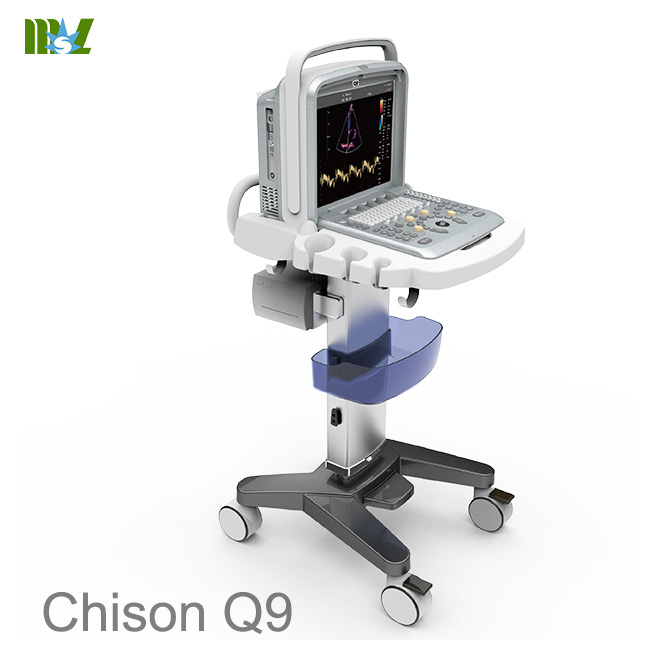
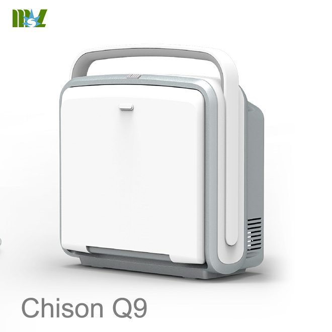
Dimensions and Weight
* Dimensions of main unit (approx.): 370mm(Length)*185mm(Width)*395mm(Height)
* Net weight of main unit (approx): 10.5kg (no probe included)
Electrical Power
* Power supply voltage: Auto adaptable for AC100V-240V
* Power supply frequency: 50-60 Hz
* Power consumption: 300 VA
Operation Panel
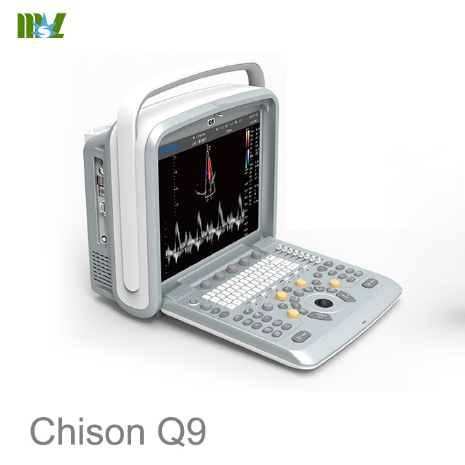
* Control panel
* Alphanumeric keyboard
* 8 TGC Slides
* Interactive backlit keys
* High resolution color LCD
- Diagonal dimension: 15 inch
- Resolution: 1024X768
- Brightness adjustment
* Integrated speaker
- Volume adjustable
Applications
* Abdomen
* Gynecology
* Obstetrics
* Urology
* Small Part
* Pediatrics
* Vascular
* Musculoskeletal
* Cardiac
Scanning Method
* Electronic convex
* Electronic linear
* Electronic micro convex
* Electronic phased array
* Volume convex
Transducer Types
* 3.5MHz Convex transducer:D3C60L
* 7.5MHz Linear transducer: D7L40L
* 7.5MHz Linear transducer: D7L60L
* 12.0MHz Linear transducer:D12L40L
* 6.0MHz Trans-vaginal transducer: D6C12L
* 7.5MHz Trans-vaginal transducer: D7C10L
* 3.0MHz Micro convex transducer:D3C20L
* 5.0MHz Micro convex transducer: D5C20L
* 6.0MHz Micro convex transducer:D6C15L
* 3.0MHz Phased array transducer: D3P64L
* 6.0MHz Phased array transducer: D6P64L
* 4.5MHz 4D volume transducer: V4C40L
Image Modes
* B mode
* 2B mode
* 4B mode
* B/M mode
* M mode
* THI mode
* B/BC mode
* CFM Mode(Color Flow Mapping)
* Pulse Doppler
* CPA (Power Doppler)
* DPD (Directional Power Doppler)
* Trapezoidal imaging (only for linear probe)
* Chroma B&M&PW
* Triplex
* Automatic PW trace and measurement in real time
* Quadplex
* Multiple Compound Imaging
* SRA( Speckle Reduction Algorithm)
* i-Image
* DICOM
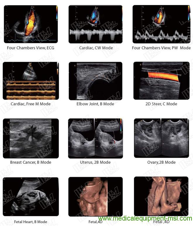
* 4D
* CWD
* Color M Mode
* ECG
* TDI
* Elastography
* Curved Panoramic Imaging
* Super needle
* Free Steering M Mode
* 2D Steer
* IMT
Display Mode
* Quad/dual display (for B, CFM , CPA)
* Duplex mode: B+CFM, B+PW, B+CPA, B+DPD, B/M
* Triplex mode: B+CFM+PW, B+CPA+PW, B+DPD+PW, B+CFM+CW, B+CPA+CW, B+DPD+CW
* Quadplex mode: B+C+PW+ Automatic PW trace and measurement in real time
Display Annotation
* Hospital name
* Date/Time
* Patient Name and Patient ID
* System status (real-time or frozen)
* Gray/Color bar
* Cine guide
* Scanning direction
* Measurement summary window
* Measurement results window
* Probe type
* Frequency
* Application name
* Menu indication
* Trackball functions indication
* Imaging parameters displayed on the screen
* TDI
* CW
Standard Configuration
* High resolution 15 inch LCD display
* 2 active transducer ports
* Pulse Wave Doppler
* Color Doppler Flow Imaging
* Power Doppler Flow Imaging
* Directional Power Doppler Flow Imaging
* ≥250G integrated hard disk
* USB ports: 2
* Ethernet port : 1
* S-video out port :1
* VGA port : 1
* General measurement package
* Clinical measurement package
* Multi-language screen display
* EASYVIEWTM: image archive system
* Patient information management system
* Building reporting system
* AIO (Automatic Image Optimization)
* Intelligent Zoom
* Speckle Reduction Algorithm (SRA)
* i-ImageTM software package
* Auto Doppler spectrum trace and auto calculate
Software Options
* DICOM 3.0: Storage, Print, Worklist, MPPS, SR Storage
* 4D software package
* Elastography
* Curved Panoramic Imaging
* Super needle
* Cardiac Package: CW, Free Steering M mode, Color M mode, TDI, ECG
* IMT
* 2D steer
Hardware Option
* 3.5MHz Convex transducer:D3C60L
* 7.5MHz Linear transducer: D7L40L
* 7.5MHz Linear transducer: D7L60L
* 12.0MHz Linear transducer: D12L40L
* 6.0MHz Trans-vaginal transducer:D6C12L
* 7.5MHz Trans-vaginal transducer: D7C10L
* 3.0MHz Micro convex transducer: D3C20L
* 5.0MHz Micro convex transducer: D5C20L
* 6.0MHz Micro convex transducer: D6C15L
* 3.0MHz Phased array transducer: D3P64L
* 6.0MHz Phased array transducer: D6P64L
* 4.5MHz 4D volume transducer: V4C40L
* CW module
* 4D module
* Biopsy kit: for convex/linear / TV probe respectively
* Footswitch
* Carrying case CS-2
* Trolley TR-2 ( Height adjustment by manual fixing )
Peripherals
* Video printer: SONY UP-897MD,SONY UP-D25MD
* PC printer :
- HP Laser Jet 1020
- HP Laser Jet CP2055d
B Mode
* Acoustic power
* Gain
* TGC
* Depth
* Freq.
* Frame rate
* Focus number
* Focus position
* Scan width
* Density
* Dynamic
* Persistence
* Noise reject
* Smooth
* Edge enhance
* i-ImageTM
* SRA
* Compound
* 2D Map
* Chroma
* Gamma
* Screen brightness
* Image rotate
* Flip (left/right, up/down)
* Zoom
* 2D steer
* Elastography
* Super needle
* Trapezoidal imaging(only for linear probe)
M Mode
* Color Map
* Sweep speed
* Layout
* Free Steering M Mode
Color Mode
* Gain
* Freq.
* Frame rate
* Steer
* PRF
* Wall filter
* Color Map
* Flow
* Color Invert
* Density
* Persistence
* Baseline
* Color mode: Velocity, Variance
* Blood Effection
* Scale
* Packet Size
* Wall Thre.
CPA/DPD Mode
* Gain
* Freq.
* Frame rate
* Steer
* PRF
* Wall filter
* Color Map
* Flow
* Density
* Persistence
* Wall Thre.
* Packet Size
PW Mode
* Gain
* PRF
* Scale
* Invert
* Wall Filter
* Audio
* Speed
* Baseline
* DA
* SV
* Color Map
* 2D Map
* Spectrum Enhance
* Dynamic Range
* Triplex
* DVmean
* DVmax
* Auto Cal
* DTrace Smooth
* Threshold
* TraceArea
CW Mode
* Gain
* PRF
* Scale
* Invert
* Wall Filter
* Audio
* Color Map
* Speed
* Baseline
* 2D Map
* CWDFFT
* CWDEnhance
* Dynamic
* DA
Cineloop
* Support 2D, M, PW, CFM, CPA, DPD, CW, Color M , Free Steering M
* Simultaneous and independent review in duplex mode
* Cineloop auto/manual
* Variable cine playback speed
* User-defined start and end frame of cine storage
* User-defined start and end frame of cine review
* Permanent storage in hard disk and display in real-time modes
* Slide show: slide show function
Storage
* ≥250GB integrated hard drive
* External USB port DVD R/W driver
* USB ports
* Still images storage format: IMAG
* Still images export format: BMP, JPG, DCM,PNG,TIFF
* Cine loops storage format: CINE
* Cine loops export format: AVI
* Fast storage setting:3s, 5s, 10s, customize time, manual
EASYVIEWTM
* Image review Layout:1×1,2×2
* Image management
- Delete selected image
- Export selected image
- Send selected image to demo
- Print selected image by PC printer
- Print selected image by DICOM printer
- Send selected image by DICOM
- Selected all
- Selected none
Exam Review
* Search Exam
* Exam review:patient view, study view
* Exam management
- Delete selected exam
- Export selected exam
- Backup selected exam
- Recover from the backup exam
- Selected all
- Expand all
- Collapse all
- Edit selected Exam
- Review selected Exam
- Continue selected Exam
General Measurement Package
- Software packages for various specific clinical use
- Comprehensive analysis methods
- Clinical analysis reports
* General measurement package
* B mode normal measurement
Distance Length_Area(Ellipse) Length_Area(Trace) Volume(1 Distance) Volume(2 Distance) Volume(3 Distance) Volume(1 Ellipse) Volume(2 Ellipse) Volume(1 Distance 1 Ellipse)
Ratio Angle
* M mode Normal measurement
MDistance MTime
Velocity Heart_Rate
* PW mode Normal measurement
Velocity Distance Peak
Auto Trace Manual Trace HR
Flow Volume StD%
StA%
Area ICA/CCA
Clinical Analysis Packages
* OB
OB -B measure Distance
Fetal Biometry: GS,CRL,YS,BPD,OFD, HC_Ellipse, APD,TAD,AC(Ellipse),FTA,FL, SL, APTD, TTD, ThC
Fetal Long Bones: Humerus,ULNA,Tibia,RAD,FIB,CLAV Fetal Cranium: CER, CM, NF, NT, OOD, IOD, NB, LVent, HW
OB Others: Lt Kid, Rt Kid, Lt Renal AP, Rt Renal AP,LV WrHEM, MAD AFI: AFI_1, AFI_2, AFI_3, AFI_4
FBP: AF
Ductus Venosus: StA%, StD%, Vessel Area, Vessel Dis StA%: A Out, A In
StD%: D Out, D In CX_L
Aorta: StA%,StD%,Veslumen_D,Veslntimal_D,VesOutside_D,Veslntimal_A,Veslumen_A StA%:A Out,A In
StD%:D Out,D In Descending Aorta:
StA%,StD%,Veslumen_D,Veslntimal_D,VesOutside_D,Veslntimal_A,Veslumen_A StA%:A Out,A In
StD%:D Out,D In MCA:
StA%,StD%, Veslumen_D,Veslntimal_D,VesOutside_D,Veslntimal_A,Veslumen_A StA%:A Out,A In
StD%:D Out,D In Umb A:
StA%,StD%, Veslumen_D,Veslntimal_D,VesOutside_D,Veslntimal_A,Veslumen_A
StA%:A Out,A In StD%:D Out,D In
Uterine Artery:Uterine Artery (Rt), Uterine Artery (Lt) Uterine Artery (Rt):
StA%,StD%, Veslumen_D,Veslntimal_D,VesOutside_D,Veslntimal_A,Veslumen_A Uterine Artery (Lt):
StA%,StD%, Veslumen_D,Veslntimal_D,VesOutside_D,Veslntimal_A,Veslumen_A StA%:A Out,A In
StD%:D Out,D In Pulmonary Artery:
StA%,StD%,Veslumen_D,Veslntimal_D,VesOutside_D,Veslntimal_A,Veslumen_A StA%:A Out,A In
StD%:D Out,D In Fetal Select
OB -D measure
Umb A Aorta
Descending Aorta Uterine Artery (Lt) Uterine Artery (Rt) Pulmonary Artery MCA
FHR
OB –M measure MDistance MTime Velocity Heart_Rate
* GYN
GYN -B measure Distance
UT:UT_L,CX_L,UT_W,UT_H
Cervix Vol.:Length,Height,Width ENDO
Right_OV_Volume:Length,Height,Width Left_OV_Volume:Length,Height,Width Right FO_D:Length,Width
Left FO_D:Length,Width
Uterine Artery:Uterine Artery(Rt),Uterine Artery(Lt),
Uterine Artery(Rt):StA%,StD%,Vessel Area,Vessel Dis Uterine Artery(Lt):StA%,StD%,Vessel Area,Vessel Dis
StA%:A Out,A In StD%:D Out,D In
GYN -D measure
Umb A:Umb A(Rt),Umb A(Lt) MCA:MCA(Rt),MCA(Lt)
Rt Uterine A:Rt Uterin A(Rt),Rt Uterin A(Lt) Lt Uterine A:Lt Uterin A(Rt),Lt Uterin A(Lt) Fetal AO:Fetal AO(Rt),Fetal AO(Lt)
FHR
GYN –M measure MDistance MTime
Velocity Heart_Rate
* Pediatrics
HIP
* URO Distance
Residual Vol. Prostate Vol. Left Kidney Right Kidney T-Zone Vol. Bladder Vol. StA%
StD%
Vessel Area Vessel Dis
* Cardiac
Cardiac-B measure Distance
Single Plane BiPlane Bullet_Volume Modi_Simpson Teichholz Cube
LV/RV AO/LV LVOT MV AV
Cardiac-D measure
LVOT AV MV TV PV
Pul.Vein HR
Cardiac-M measure Distance Heart_Rate Ejection_Time
LV LVSHORT AV AVSHORT MV
AV AO/LV LVOT TV
PulV
* Vessel IMT(Auto)
Prox CCA Mid CCA Distal CCA
Prox ICA Mid ICA Distal ICA ECA
Vertebral A INT IIL EXT IL ILIAC
CFA
ProFun LTCIR SFA
Pop A ATA PTA PERON DRPED
* Abdomen CBD
GB Wall Liver Length Prox Aorta Mid Aorta Distal Aorta Spleen Renal Vol. Lliac
* Carotid Subclavian A Prox CCA Mid CCA Distal CCA Bulb
Prox ICA Mid ICA Distal ICA ECA
Vertebral A
General Measurement Flow Volume
* Small parts
General Measurement Ratio
Angle
By using system setup, users could
* Customize hospital information
* Customize language
* Customize fast storage time
* Customize color map
* Assign functions to “PRINT” button on control panel and foot switch
* Customize comment library
* Customize report
User Define Functions
By user-define function, users could customize user-define preset, including
- Applications name, Presets name, User defined name
- Applications exam type
- Imaging parameters
Multi-language Display Interface
* English
* Chinese
* Polish
* Portuguese
* Russian
* Spanish
* Danish
* German
* French
Operation System
Windows XP Embedded
* S-video: 1
* Video out: 1
* VGA: 1
* USB port: 2
* Ethernet: 1
* Remote control: 1
* Footswitch port: 1
* System power in: 1
* Ground pole: 1
* Power button: 1
* Ambient temperature: 10°C to 40°C
* Relative humidity: 30% to 75%
* Atmospheric pressure: 700 hPa to 1060 hPa
* Ambient temperature: -5°C to 40°C
* Relative humidity: ≤80% (no condensation)
* Atmospheric pressure: 700 hPa to 1060 hPa
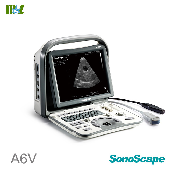
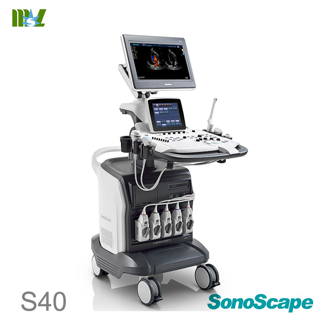
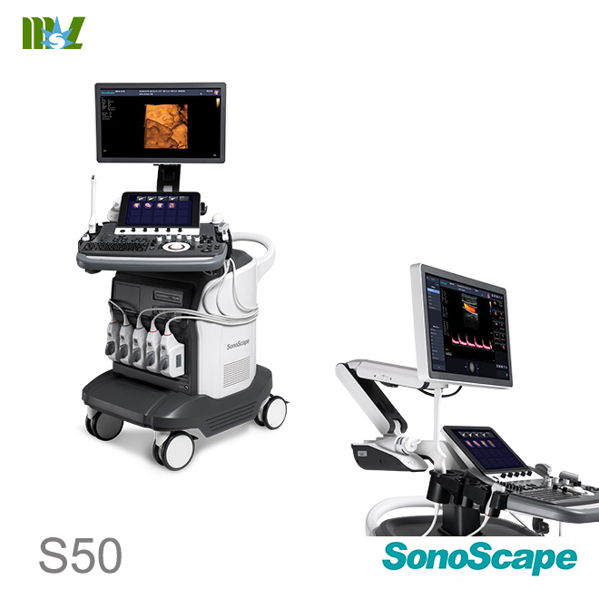
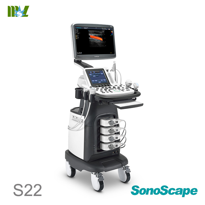
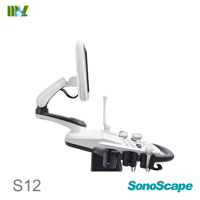
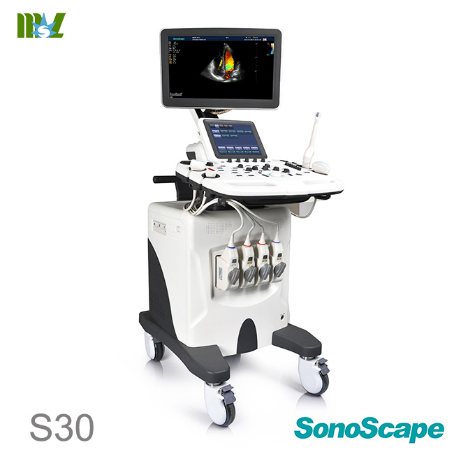
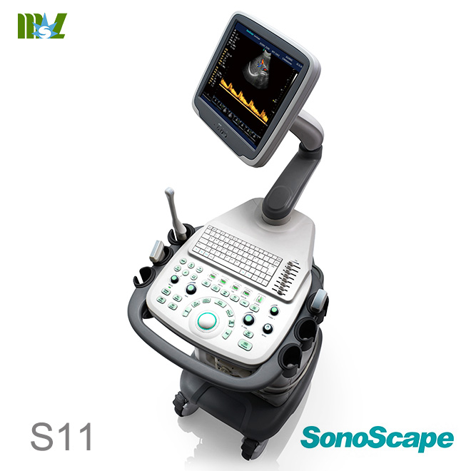
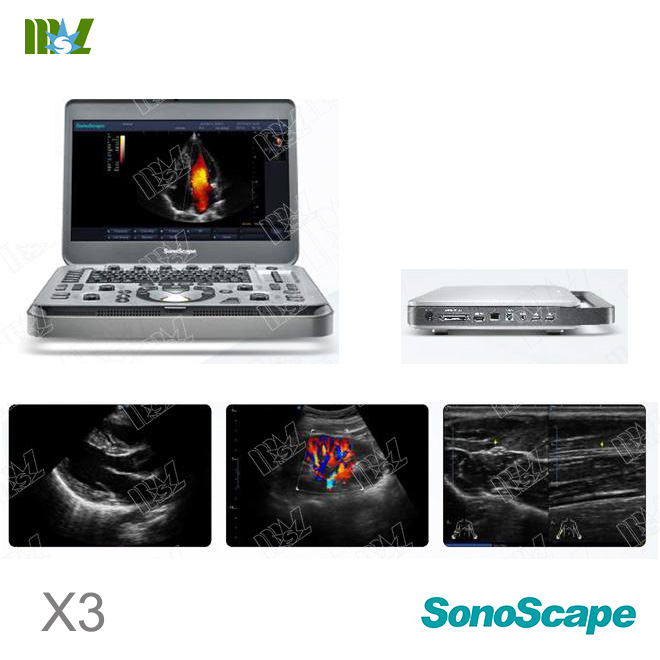
Hot sale Sonoscape ultrasound | Chison ultrasound price list
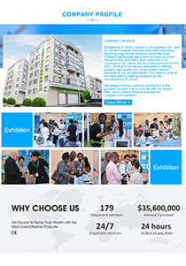
MSL TEAM picture


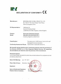
MSL Certificate
MSL Medical cooperate with DHL,FEDEX,UPS,EMS,TNT,etc.International shipping company,make your goods arrive destination safely and quickly.


 Price is 8-20% Lower Than Other



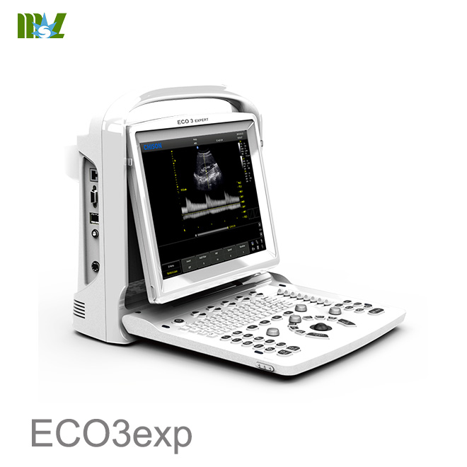
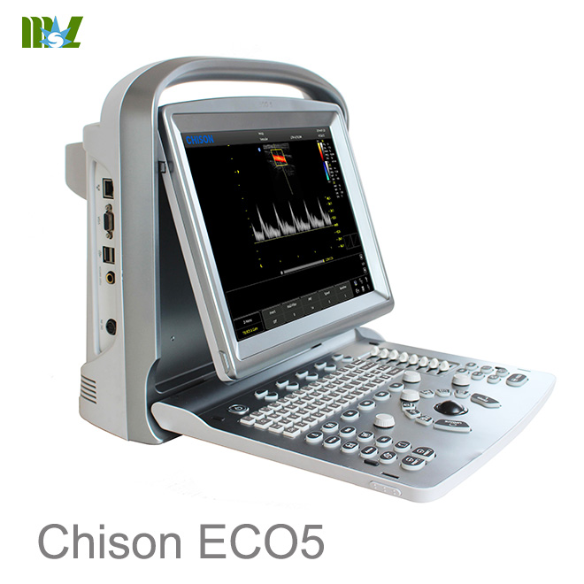
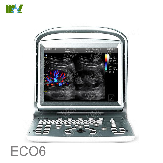


![{pr0int $v['title']/}](https://medicalequipment-msl.com/upload/img/20170705/201707051810527163.jpg.jpg)
![{pr0int $v['title']/}](https://medicalequipment-msl.com/upload/img/20170706/201707061632154149.jpg.jpg)
![{pr0int $v['title']/}](https://medicalequipment-msl.com/upload/img/20170706/201707061715033181.jpg.jpg)
![{pr0int $v['title']/}](https://medicalequipment-msl.com/upload/img/20220318/202203181130316800.jpg.jpg)
![{pr0int $v['title']/}](https://medicalequipment-msl.com/upload/img/20170706/201707061150472678.jpg.jpg)
![{pr0int $v['title']/}](https://medicalequipment-msl.com/upload/img/20170706/201707061726005808.jpg.jpg)
![{pr0int $v['title']/}](https://medicalequipment-msl.com/upload/img/20170706/201707061532054748.jpg.jpg)
![{pr0int $v['title']/}](https://medicalequipment-msl.com/upload/img/20180511/201805111718487852.jpg.jpg)
![{pr0int $v['title']/}](https://medicalequipment-msl.com/upload/img/20170706/201707061746321676.jpg.jpg)
![{pr0int $v['title']/}](https://medicalequipment-msl.com/upload/img/20170706/201707061522373256.jpg.jpg)
![{pr0int $v['title']/}](https://medicalequipment-msl.com/upload/img/20210803/202108031117167933.jpg.jpg)
![{pr0int $v['title']/}](https://medicalequipment-msl.com/upload/img/20210803/202108031127169036.jpg.jpg)
![{pr0int $v['title']/}](https://medicalequipment-msl.com/upload/img/20210803/20210803115002472.jpg.jpg)
![{pr0int $v['title']/}](https://medicalequipment-msl.com/upload/img/20210803/202108031150022887.jpg.jpg)
![{pr0int $v['title']/}](https://medicalequipment-msl.com/upload/img/20210803/202108031415035491.jpg.jpg)
![{pr0int $v['title']/}](https://medicalequipment-msl.com/upload/img/20210803/202108031415038949.jpg.jpg)
![{pr0int $v['title']/}](https://medicalequipment-msl.com/upload/img/20210803/202108031431393763.jpg.jpg)
![{pr0int $v['title']/}](https://medicalequipment-msl.com/upload/img/20210803/202108031438195289.jpg.jpg)
![{pr0int $v['title']/}](https://medicalequipment-msl.com/upload/img/20210803/202108031447282738.jpg.jpg)
![{pr0int $v['title']/}](https://medicalequipment-msl.com/upload/img/20210804/202108040929504520.jpg.jpg)
![{pr0int $v['title']/}](https://medicalequipment-msl.com/upload/img/20210804/202108041023201613.jpg.jpg)
![{pr0int $v['title']/}](https://medicalequipment-msl.com/upload/img/20210804/202108041021051696.jpg.jpg)
![{pr0int $v['title']/}](https://medicalequipment-msl.com/upload/img/20210804/202108041448486487.jpg.jpg)
![{pr0int $v['title']/}](https://medicalequipment-msl.com/upload/img/20210804/202108041432386584.jpg.jpg)
![{pr0int $v['title']/}](https://medicalequipment-msl.com/upload/img/20210804/202108041432385602.jpg.jpg)


