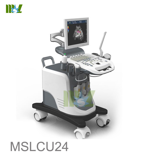
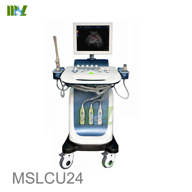
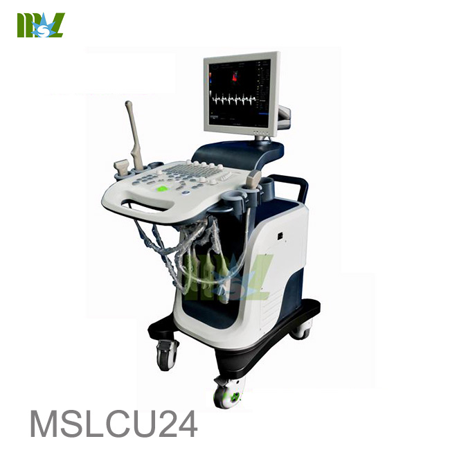
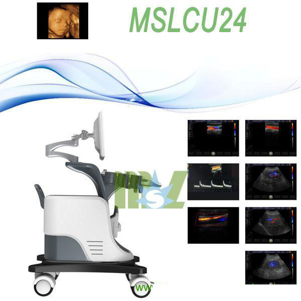
MSL 4D Color Doppler Ultrasound Machine / 4D Baby Ultrasound pregnancy MSLCU24 for sale
*4D ultrasound machine pregnancy / 4d USG
*Trolley ultrasound machine
*Full digital beam forming technology
*15 inch LCD color display
*Running hours: ≥8h
*Input power: ≤320V
4D Baby Ultrasound pregnancy MSLCU24 Main Functions and Technical Indexes
MSL 4D Color Doppler Ultrasound Machine / 4D Baby Ultrasound pregnancy MSLCU24 for sale
YOUTUBE video link:
MSL 4D Baby Ultrasound pregnancy MSLCU24 /4D Color Doppler video
VIDEO
MSL 4D Color Doppler Ultrasound Machine operation Video
VIDEO
MSL 4D Color Doppler Ultrasound Machine pregnacy MSLCU24 Operating environment
Operating ambient temperature:+5℃~+40℃
Relative humidity:≤80%
Atmospheric pressure: 700hPa—1060hPa
Power supply:AC220V±22V,50Hz±1Hz
Plug seat shall have independent power supply.
Be far away from strong electric field, strong magnetic field equipment and high voltage equipment.
4D Baby Ultrasound pregnancy MSLCU24 Main Functions
Having a full digital beam forming technology
Scanning mode: Convex array, lumen, high-frequency linear array, phased array, 3D software(Optional accessories);
Dynamic range: 0~120dB adjustable;
Display mode: B、B/B、M、B/M、CFM、CMF/B、PDI、B/PW, total eight mode;
Application mode: abdomen, gynecology, obstetrics, superficial organ, urologist, heart and user defined model 1-4, total ten models;
Image mode: digital beam forming, tissue harmonic imaging;
Acoustic output: Mechanical index and thermal index real-time display;
Acoustic power : Step is adjustable, real-time display;
Gray scale: 256 scales;
Depth display: ≥250mm;
B/D dual-purpose: linear array: B/PWD; convex array: B/PWD;
Pseudo color processing: 16 kinds of pseudo color encoding can optional;
Gain adjusts: 8 segments TGC, B/M/D/C is independently adjustable; TGC curve can show and hide automatically;
Image magnification: picture in picture zoom in and zoom part function;
Image processing: Edge enhancement: Multilevel adjustable
Frame average: Multilevel adjustable
Line average: Multilevel adjustable
Focus Optimization: Multilevel adjustable
Gray Restrain: Multilevel adjustable
Gamma correction: Multilevel adjustable
Contrast: Adjustable
Brightness: Adjustable
Self-motion optimize function: Built-in multiple check type, according to different inspection organs, preset best image check condition, reduce the adjusting operation keys;
One-click optimization function: preset several parameters adjusting focus on a button, a key to realize image fast optimization;
Measurement and calculation:
B mode routine measurement:
Distance, circumference, area, volume, angle, ratio, and stenos rate.
M mode routine measurement: Heart rate, time, distance, speed, ratio, etc..
Gynecology measurement: Uterus, cervix, endometrial, ovary, follicular.
Obstetrics measurement:
EGA, ETD, fetal weight estimation, AFI index, OB report (including OB tables).
Cardiology measurement: LV measurement.
Urology measurement:
Prostate volume, displacement volume, bladder capacity, and residual urine output.
PW measurements: Time, speed, Heart Rate, RI, PI, etc.
Other measurement: Slice volume measurement, hip joint angle measurement.
Image storage: Image storage, video storage, cine loop, disk storage capacity≥160G;
Patient data: Medical record management, report inquiry and printing, image video output( HDD 、DVD-RW、USB)、built-in ultrasound workstation;
Reporting system: automatic report generation system, and can be full screen characters in both Chinese and English editor;
Output interface: SR323、USB、DICOM interface;
4D Baby Ultrasound pregnancy MSLCU24 Main Technical Indexes
1.The performance requirements of gray-scale imaging mode
The color ultrasonic at the gray-scale imaging performance mode should comply with the provisions of the table 1.
Table 1 At the Gray-scale imaging mode the performance of the probe
performance indexes
probe type and nominal frequency
2.0≤f<4.0
2.0≤f<5.0
5.0≤f<8.0
5.0≤f<10.0
a) probe type and model
phased array (type TP16A)
Convex array
(type TC60A)
Cavity
(type TC10A)
Linear array
(type TL40A)
b) nominal frequency (MHz)
3.0
3.5
6.5
7.5
c) Scan depth(mm)
≥140
≥160
≥40
≥50
d) Lateral resolution (mm)
≤3(depth≤80)
≤4(80<depth≤130)
≤3(depth≤80)
≤4(80<depth≤130)
≤2(depth≤30)
≤2(depth≤40)
e) Axial resolution (mm)
≤2(depth≤80)
≤2(depth≤80)
≤3(80<depth≤130)
≤1(depth≤40)
≤1(depth≤50)
f) Blind area (mm)
≤7
≤5
≤4
≤3
g) Transverse geometry precision (%)
≤20
≤15
≤10
≤10
h) Longitudinal geometric location accuracy (%)
≤10
≤10
≤5
≤5
i) Slice thickness (mm)
≤5
≤5
≤5
≤5
j) Perimeter and area measured deviation (%)
≤±20
≤±20
≤±20
≤±20
k) M mode time display error (%)
≤±10
≤±10
≤±10
≤±10
2 .The performance requirements of color Doppler imaging mode
a). The color ultrasonic at the color Doppler imaging mode should comply with the provisions of the
table 2.2;
b). Color blood flow image should be essentially coincident with the gray-scale image of pipe’s;
c). Blood flow direction should be able to correctly identify, no aliasing phenomenon;
Table 2 at the color blood flow imaging mode the performance of the probe
Doppler model
Phased array
Convex array
Cavity
Linear array
Investigation depth at Color blood flow model
≥90mm
≥100mm
≥40mm
≥50mm
Investigation depth at Doppler spectrum model
≥90mm
≥100mm
≥40mm
≥50mm
Blood flow speed reading error
≤±15%
3 The performance requirements of Doppler spectrum mode
a). The color ultrasonic at the color Doppler spectrum mode should comply with the provisions of the;
b). Blood flow speed reading error should comply with the provisions of the table2.3;
c). Pulse wave Doppler mode sampling area cursor position should be accurate;
Table 3 at the color blood flow imaging mode the performance of the probe
Doppler model
Phased array
Convex array
Cavity
Linear array
Investigation depth at Color blood flow model
≥90mm
≥100mm
≥40mm
≥50mm
Investigation depth at Doppler spectrum model
≥90mm
≥100mm
≥40mm
≥50mm
Blood flow speed reading error
≤±15%
Displayer: 15 inch LCD color display
Running hours: ≥8h;
Input power: ≤320V;
Host weight: about 75 kg;
Host appearance size: 1140 ×690× 1070(length × width × height) (mm 3 )
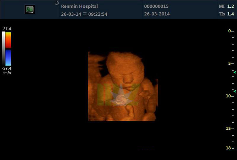
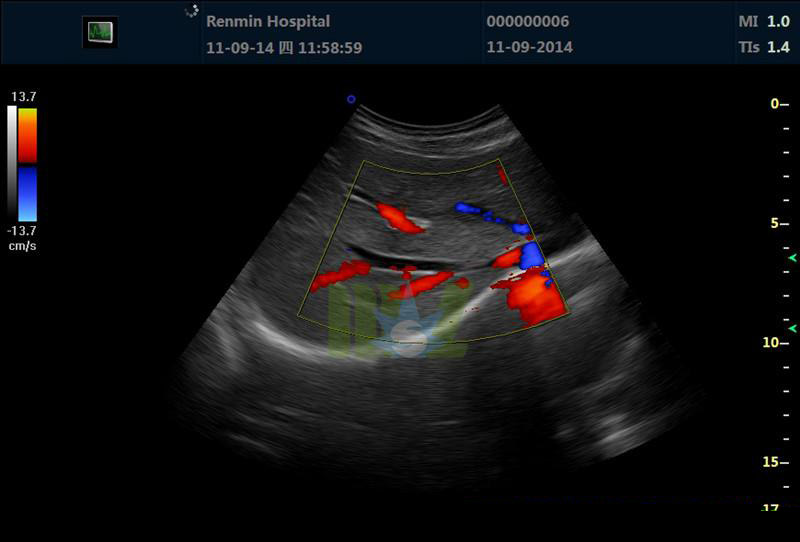
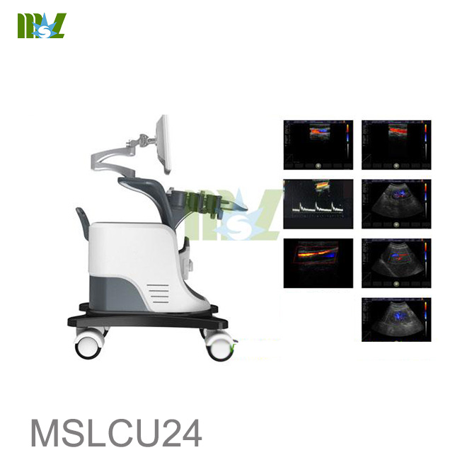
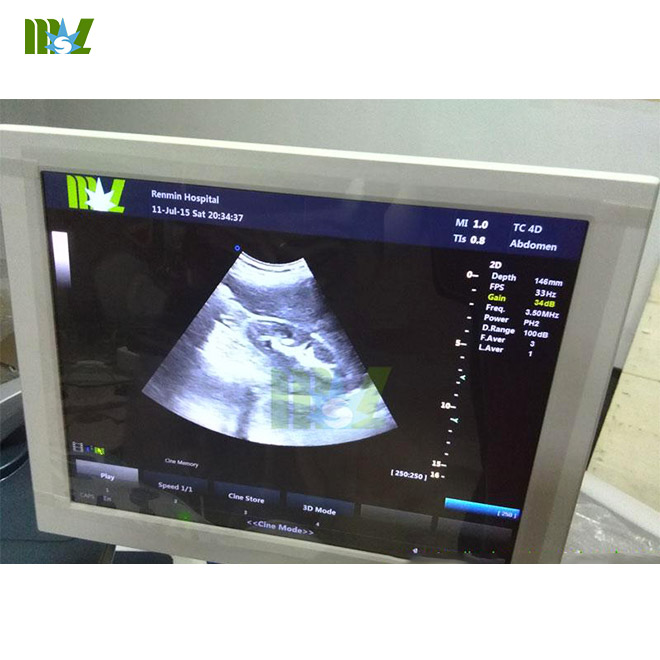
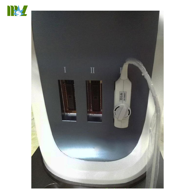
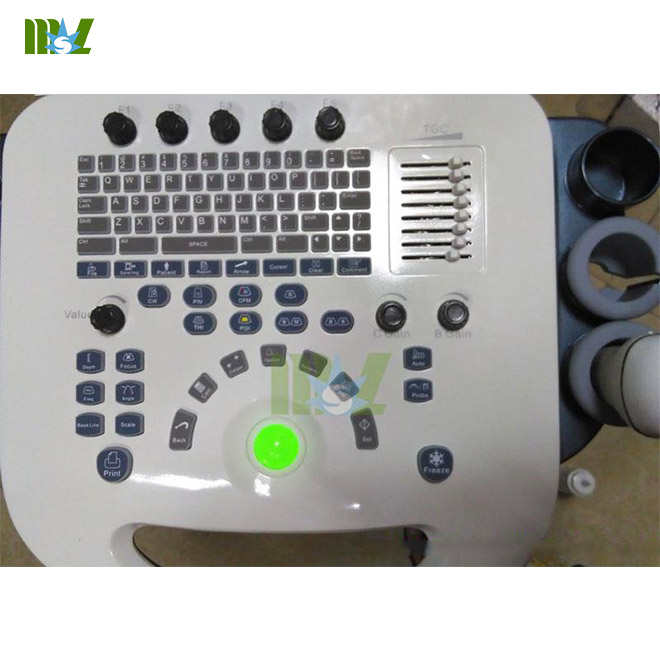
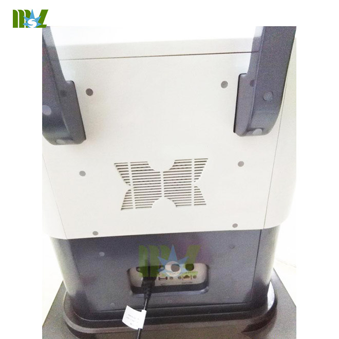
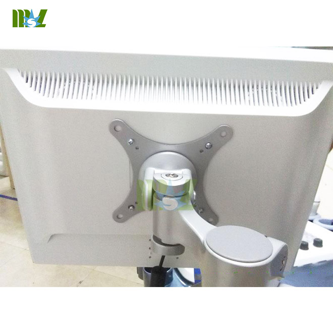
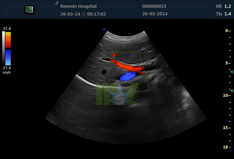
Effect pictures of the 4D Baby Ultrasound pregnancy MSLCU24
what will you get?
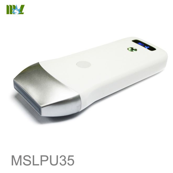
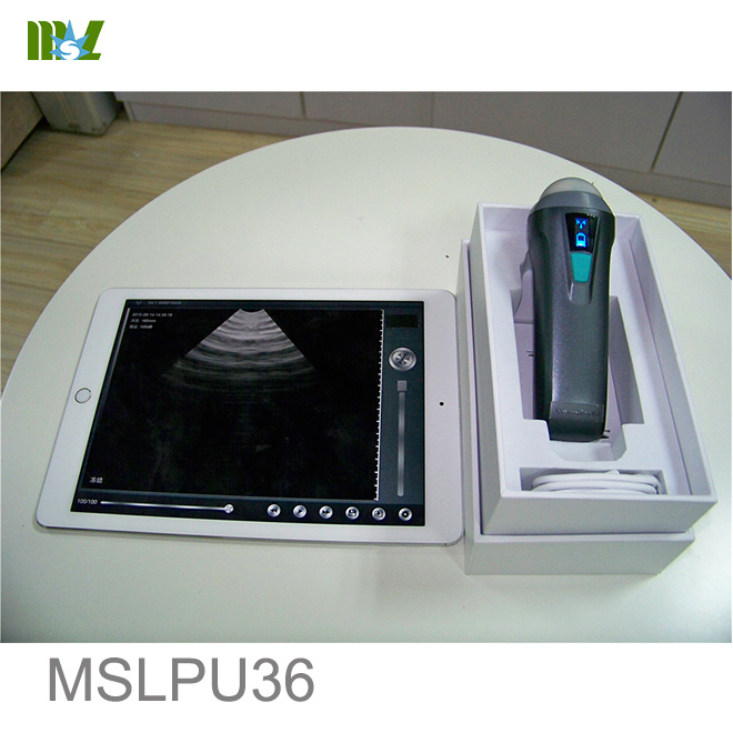
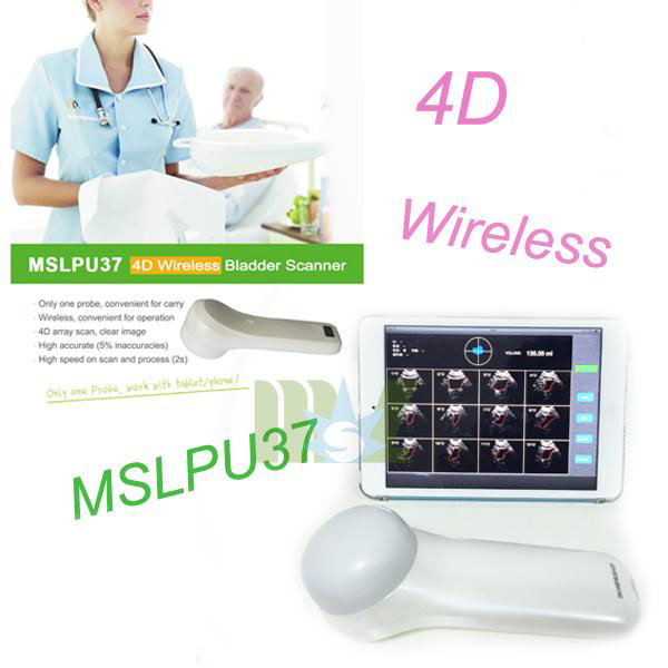
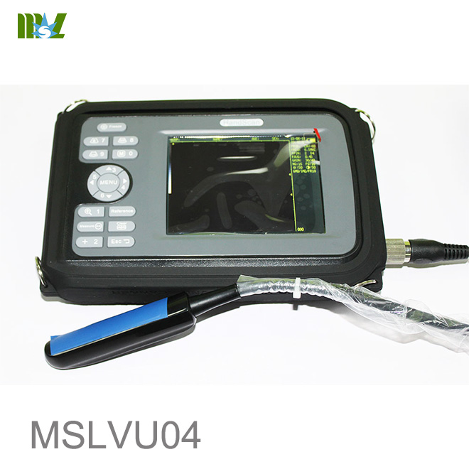
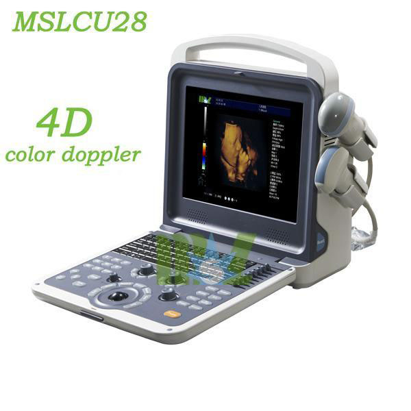
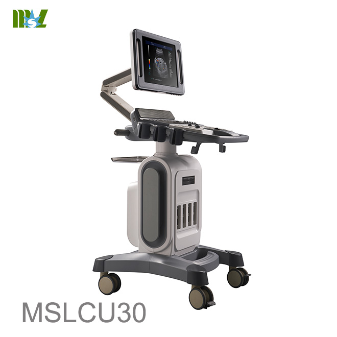
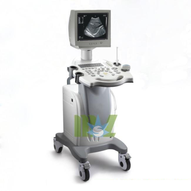
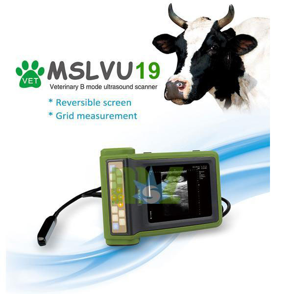
Other Hot sale ultrasound machine recommend
MSL Certificate
MSL Medical cooperate with DHL,FEDEX,UPS,EMS,TNT,etc.International shipping company,make your goods arrive destination safely and quickly.


 Price is 8-20% Lower Than Other



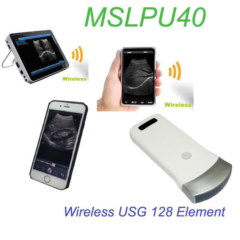
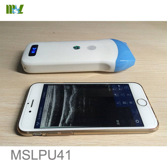
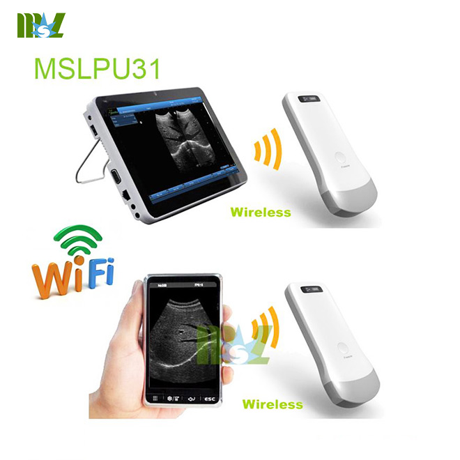



![{pr0int $v['title']/}](https://medicalequipment-msl.com/upload/img/20150505/201505050942035656.jpg.jpg)
![{pr0int $v['title']/}](https://medicalequipment-msl.com/upload/img/20150820/201508201430246682.jpg.jpg)
![{pr0int $v['title']/}](https://medicalequipment-msl.com/upload/img/20160226/201602261629209261.jpg.jpg)
![{pr0int $v['title']/}](https://medicalequipment-msl.com/upload/img/20170928/201709281055391381.jpg.jpg)
![{pr0int $v['title']/}](https://medicalequipment-msl.com/upload/img/20170927/201709271820337067.jpg.jpg)
![{pr0int $v['title']/}](https://medicalequipment-msl.com/upload/img/20170928/20170928114415198.jpg.jpg)
![{pr0int $v['title']/}](https://medicalequipment-msl.com/upload/img/20211222/202112221706187921.jpg.jpg)
![{pr0int $v['title']/}](https://medicalequipment-msl.com/upload/img/20160331/201603311420026640.jpg.jpg)
![{pr0int $v['title']/}](https://medicalequipment-msl.com/upload/img/20221020/202210201655209109.jpg.jpg)


